JUN MA
junma
- Canada
- University of Toronto
- Laboratory Medicine and Pathobiology
- Website
Statistics
- Member for 6 years, 2 months
- 136 challenge submissions
- 3 algorithms run
Activity Overview
PAVES
Challenge UserPeripheral Artery:Vein Enhanced Segmentation (PAVES) is the challenge focussed on providing easily interpretable and relevant images that can be readily understood by clinicians (vascular interventional radiologists & vascular surgeons) from MRA datasets where the venous and arterial vasculature may be equally enhanced. The setting is lower limb arterial occlusive disease where imaging of the below knee arterial vasculature is critical in planning limb salvage interventions. However, the competing demands of the high spatial resolution needed to image small vessels versus imaging time constraints where there is often a very short arteriovenous transit time for contrast passage form arterial to venous compartments makes imaging challenging. While dynamic MRA techniques can usually allow arterial imaging without venous ‘contamination’ these necessarily sacrifice spatial resolution.


PAIP2020
Challenge UserBuilt on the success of its predecessor, PAIP2020 is the second challenge organized by the Pathology AI Platform (PAIP) and the Seoul National University Hospital (SNUH). PAIP2020 will proceed to not only detect whole tumor areas in colorectal cancers but also to classify their molecular subtypes, which will lead to characterization of their heterogeneity with respect to prognoses and therapeutic responses. All participants should predict one of the molecular carcinogenesis pathways, i.e., microsatellite instability(MSI) in colorectal cancer, by performing digital image analysis without clinical tests. This task has a high clinical relevance as the currently used procedure requires an extensive microscopic assessment by pathologists. Therefore, those automated algorithms would reduce the workload of pathologists as a diagnostic assistance.
Thyroid Nodule Segmentation and Classification
Challenge UserThe main topic of this TN-SCUI2020 challenge is finding automatic algorithms to accurately classify the thyroid nodules in ultrasound images. It will provide the biggest public dataset of thyroid nodule with over 4500 patient cases from different ages, genders, and were collected using different ultrasound machines. Each ultrasound image is provided with its ground truth class (benign or maglinant) and a detailed delineation of the nodule. This challenge will provide a unique opportunity for participants from different backgrounds (e.g. academia, industry, and government, etc.) to compare their algorithms in an impartial way.
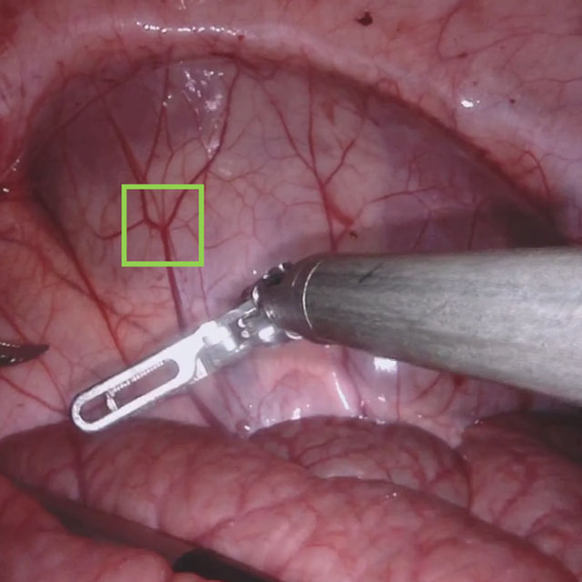
SARAS-ESAD
Challenge UserThis challenge is part of Medical Imaging with Deep Learning conference, 2020. The conference is held between 6 ‑ 8 July 2020 in Montréal. The SARAS (Smart Autonomous Robotic Assistant Surgeon) EU consortium, www.saras-project.eu, is working towards replacing the assistant surgeon in MIS with two assistive robotic arms. To accomplish that, an artificial intelligence based system is required which not only can understand the complete surgical scene but also detect the actions being performed by the main surgeon. This information can later be used infer the response required from the autonomous assistant surgeon.

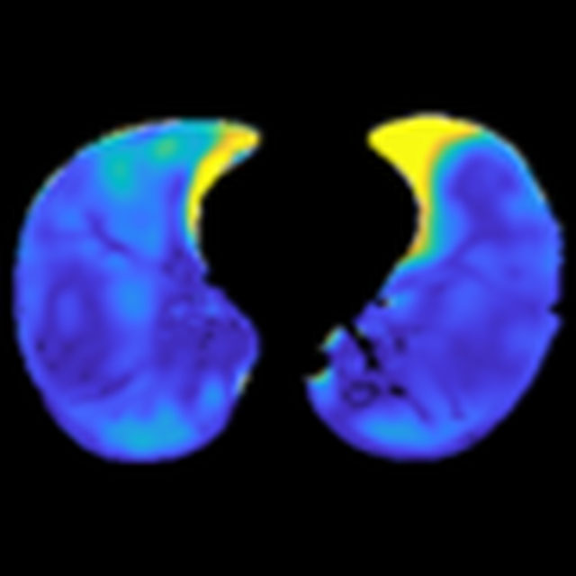
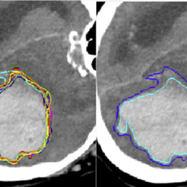
CADA
Challenge UserCerebral aneurysms are local dilations of arterial blood vessels caused by a weakness of the vessel wall. Subarachnoid hemorrhage (SAH) caused by the rupture of a cerebral aneurysm is a life-threatening condition associated with high mortality and morbidity. The mortality rate is above 40%, and even in case of survival cognitive impairment can affect patients for a long time. Major goals in image analysis are the detection and risk assessment of aneurysms. We, therefore, subdivided the challenge into three categories. The first task is finding the aneurysm; the second task is the accurate segmentation to allow for a longitudinal assessment of the development of suspicious aneurysms. The third task is the estimation of the rupture risk of the aneurysm.


CADA - Rupture Risk Estimation
Challenge UserCerebral aneurysms are local dilations of arterial blood vessels caused by a weakness of the vessel wall. Subarachnoid hemorrhage (SAH) caused by the rupture of a cerebral aneurysm is a life-threatening condition associated with high mortality and morbidity. The mortality rate is above 40%, and even in case of survival cognitive impairment can affect patients for a long time. Major goals in image analysis are the detection and risk assessment of aneurysms. We, therefore, subdivided the challenge into three categories. The first task is finding the aneurysm; the second task is the accurate segmentation to allow for a longitudinal assessment of the development of suspicious aneurysms. The third task is the estimation of the rupture risk of the aneurysm.
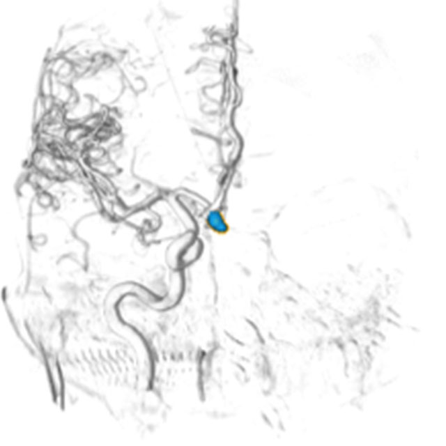
CADA - Aneurysm Segmentation
Challenge UserCerebral aneurysms are local dilations of arterial blood vessels caused by a weakness of the vessel wall. Subarachnoid hemorrhage (SAH) caused by the rupture of a cerebral aneurysm is a life-threatening condition associated with high mortality and morbidity. The mortality rate is above 40%, and even in case of survival cognitive impairment can affect patients for a long time. Major goals in image analysis are the detection and risk assessment of aneurysms. We, therefore, subdivided the challenge into three categories. The first task is finding the aneurysm; the second task is the accurate segmentation to allow for a longitudinal assessment of the development of suspicious aneurysms. The third task is the estimation of the rupture risk of the aneurysm.

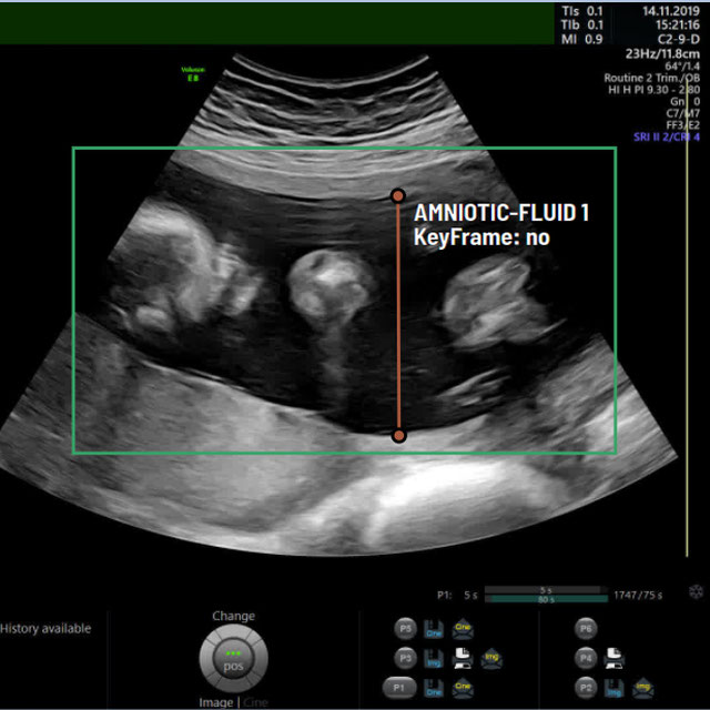
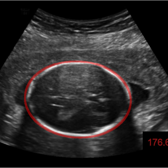
A-AFMA
Challenge UserPrenatal ultrasound (US) measurement of amniotic fluid is an important part of fetal surveillance as it provides a non-invasive way of assessing whether there is oligohydramnios (insufficient amniotic fluid) and polyhydramnios (excess amniotic fluid), which are associated with numerous problems both during pregnancy and after birth. In this Image Analysis Challenge, we aim to attract attention from the image analysis community to work on the problem of automated measurement of the MVP using the predefined ultrasound video clip based on a linear-sweep protocol [1]. We define two tasks. The first task is to automatically detect amniotic fluid and the maternal bladder. The second task is to identify the appropriate points for MVP measurement given the selected frame of the video clip, and calculate the length of the connected line between these points. The data was collected from women in the second trimester of pregnancy, as part of the PURE study at the John Radcliffe Hospital in Oxford, UK.


WSSS4LUAD
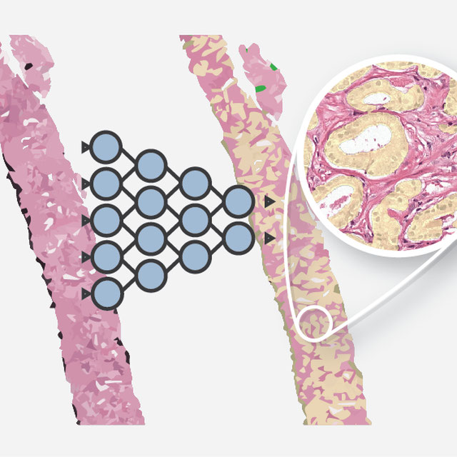
Challenge UserThe WSSS4LUAD dataset contains over 10,000 patches of lung adenocarcinoma from whole slide images from Guangdong Provincial People's Hospital and TCGA with image-level annotations. The goal of this challenge is to perform semantic segmentation for differentiating three important types of tissues in the WSIs of lung adenocarcinoma, including cancerous epithelial region, cancerous stroma region and normal region. Paticipants have to use image-level annotations to give pixel-level prediction.

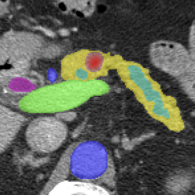
Parse2022
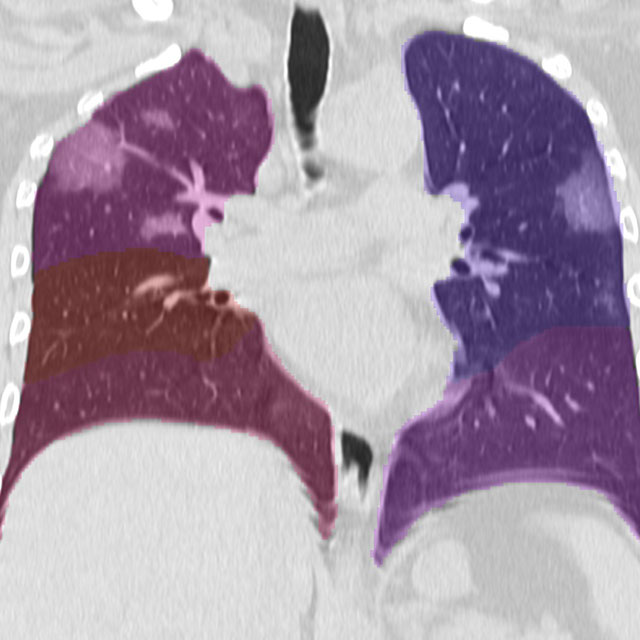
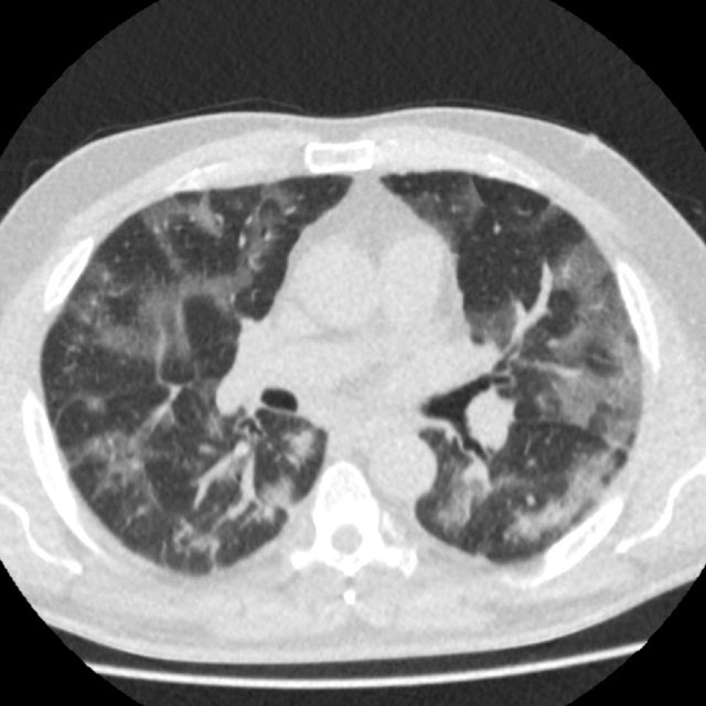
Challenge UserIt is of significant clinical interest to study pulmonary artery structures in the field of medical image analysis. One prerequisite step is to segment pulmonary artery structures from CT with high accuracy and low time-consuming. The segmentation of pulmonary artery structures benefits the quantification of its morphological changes for diagnosis of pulmonary hypertension and thoracic surgery. However, due to the complexity of pulmonary artery topology, automated segmentation of pulmonary artery topology is a challenging task. Besides, the open accessible large-scale CT data with well labeled pulmonary artery are scarce (The large variations of the topological structures from different patients make the annotation an extremely challenging process). The lack of well labeled pulmonary artery hinders the development of automatic pulmonary artery segmentation algorithm. Hence, we try to host the first Pulmonary ARtery SEgmentation challenge in MICCAI 2022 (Named Parse2022) to start a new research topic.
3D Teeth Scan Segmentation and Labeling Challenge MICCAI2022
Challenge UserComputer-aided design (CAD) tools have become increasingly popular in modern dentistry for highly accurate treatment planning. In particular, in orthodontic CAD systems, advanced intraoral scanners (IOSs) are now widely used as they provide precise digital surface models of the dentition. Such models can dramatically help dentists simulate teeth extraction, move, deletion, and rearrangement and therefore ease the prediction of treatment outcomes. Although IOSs are becoming widespread in clinical dental practice, there are only few contributions on teeth segmentation/labeling available in the literature and no publicly available database. A fundamental issue that appears with IOS data is the ability to reliably segment and identify teeth in scanned observations. Teeth segmentation and labelling is difficult as a result of the inherent similarities between teeth shapes as well as their ambiguous positions on jaws.


ToothFairy: Cone-Beam Computed Tomography Segmentation Challenge
Challenge UserThis is the first edition of the ToothFairy challenge organized by the University of Modena and Reggio Emilia with the collaboration of Raudboud University. This challenge aims at pushing the development of deep learning frameworks to segment the Inferior Alveolar Canal (IAC) by incrementally extending the amount of publicly available 3D-annotated Cone Beam Computed Tomography (CBCT) scans. CBCT modality is becoming increasingly important for treatment planning and diagnosis in implant dentistry and maxillofacial surgery. The three-dimensional information acquired with CBCT can be crucial to plan a vast number of surgical interventions with the aim of preserving noble anatomical structures such as the Inferior Alveolar Canal (IAC), which contains the homonymous nerve (Inferior Alveolar Nerve, IAN). Deep learning models can support medical personnel in surgical planning procedures by providing a voxel-level segmentation of the IAN automatically extracted from CBCT scans.

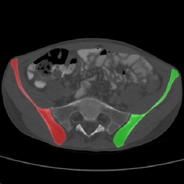
LNQ2023
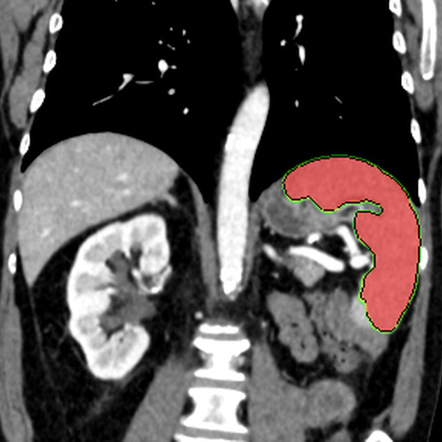
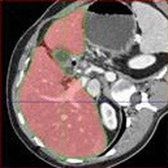
Challenge UserAccurate lymph node size estimation is critical for staging cancer patients, initial therapeutic management, and in longitudinal scans, assessing response to therapy. Current standard practice for quantifying lymph node size is based on a variety of criteria that use unidirectional or bidirectional measurements on just one or a few nodes, typically on just one axial slice. But humans have hundreds of lymph nodes, any number of which may be enlarged to various degrees due to disease or immune response. While a normal lymph node may be approximately 5mm in diameter, a diseased lymph node may be several cm in diameter. The mediastinum, the anatomical area between the lungs and around the heart, may contain ten or more lymph nodes, often with three or more enlarged greater than 1cm. Accurate segmentation in 3D would provide more information to evaluate lymph node disease.
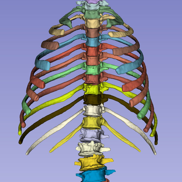
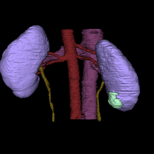
SEG.A. - Segmentation of the Aorta
Challenge UserSegmentation, modeling and visualization of the arterial tree are still a challenge in medical image analysis. The main track of this challenge deals with the fully automatic segmentation of the aortic vessel tree in computed tomography images. Optionally, teams can submit tailored solutions for meshing and visualization of the vessel tree.


Light My Cells : Bright Field to Fluorescence Imaging Challenge
Challenge UserJoin the Light My Cells France-Bioimaging challenge! Enhance biology and microscopy by contributing to the development of new image-to-image deep labelling methods. The task: predict the best-focused output images of several fluorescently labelled organelles from label-free transmitted light input images. Dive into the future of imaging with us! 🌐🔬 #LightMyCellsChallenge

AI4Life Microscopy Denoising Challenge
Challenge UserWellcome to AI4Life-MDC24! In this challenge, we want to focus on an unsupervised denoising of microscopy images. By participating, researchers can contribute to a critical area of scientific research, aiding in interpreting microscopy images and potentially unlocking discoveries in biology and medicine.