Kele XU
kele
- France
- Langevin Institute
- Engineering
Statistics
- Member for 8 years, 2 months
- 31 challenge submissions
Activity Overview

Thyroid Nodule Segmentation and Classification
Challenge UserThe main topic of this TN-SCUI2020 challenge is finding automatic algorithms to accurately classify the thyroid nodules in ultrasound images. It will provide the biggest public dataset of thyroid nodule with over 4500 patient cases from different ages, genders, and were collected using different ultrasound machines. Each ultrasound image is provided with its ground truth class (benign or maglinant) and a detailed delineation of the nodule. This challenge will provide a unique opportunity for participants from different backgrounds (e.g. academia, industry, and government, etc.) to compare their algorithms in an impartial way.
SARAS-ESAD
Challenge UserThis challenge is part of Medical Imaging with Deep Learning conference, 2020. The conference is held between 6 ‑ 8 July 2020 in Montréal. The SARAS (Smart Autonomous Robotic Assistant Surgeon) EU consortium, www.saras-project.eu, is working towards replacing the assistant surgeon in MIS with two assistive robotic arms. To accomplish that, an artificial intelligence based system is required which not only can understand the complete surgical scene but also detect the actions being performed by the main surgeon. This information can later be used infer the response required from the autonomous assistant surgeon.

CADA
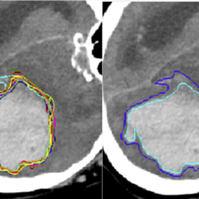
Challenge UserCerebral aneurysms are local dilations of arterial blood vessels caused by a weakness of the vessel wall. Subarachnoid hemorrhage (SAH) caused by the rupture of a cerebral aneurysm is a life-threatening condition associated with high mortality and morbidity. The mortality rate is above 40%, and even in case of survival cognitive impairment can affect patients for a long time. Major goals in image analysis are the detection and risk assessment of aneurysms. We, therefore, subdivided the challenge into three categories. The first task is finding the aneurysm; the second task is the accurate segmentation to allow for a longitudinal assessment of the development of suspicious aneurysms. The third task is the estimation of the rupture risk of the aneurysm.

CT diagnosis of COVID-19
Challenge UserCoronavirus disease 2019 (COVID-19) has infected more than 1.3 million individuals all over the world and caused more than 106,000 deaths. One major hurdle in controlling the spreading of this disease is the inefficiency and shortage of medical tests. To mitigate the inefficiency and shortage of existing tests for COVID-19, we propose this competition to encourage the development of effective Deep Learning techniques to diagnose COVID-19 based on CT images. The problem we want to solve is to classify each CT image into positive COVID-19 (the image has clinical findings of COVID-19) or negative COVID-19 ( the image does not have clinical findings of COVID-19). It’s a binary classification problem based on CT images.

CADA - Rupture Risk Estimation
Challenge UserCerebral aneurysms are local dilations of arterial blood vessels caused by a weakness of the vessel wall. Subarachnoid hemorrhage (SAH) caused by the rupture of a cerebral aneurysm is a life-threatening condition associated with high mortality and morbidity. The mortality rate is above 40%, and even in case of survival cognitive impairment can affect patients for a long time. Major goals in image analysis are the detection and risk assessment of aneurysms. We, therefore, subdivided the challenge into three categories. The first task is finding the aneurysm; the second task is the accurate segmentation to allow for a longitudinal assessment of the development of suspicious aneurysms. The third task is the estimation of the rupture risk of the aneurysm.
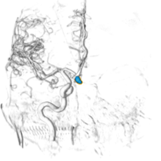
CADA - Aneurysm Segmentation
Challenge UserCerebral aneurysms are local dilations of arterial blood vessels caused by a weakness of the vessel wall. Subarachnoid hemorrhage (SAH) caused by the rupture of a cerebral aneurysm is a life-threatening condition associated with high mortality and morbidity. The mortality rate is above 40%, and even in case of survival cognitive impairment can affect patients for a long time. Major goals in image analysis are the detection and risk assessment of aneurysms. We, therefore, subdivided the challenge into three categories. The first task is finding the aneurysm; the second task is the accurate segmentation to allow for a longitudinal assessment of the development of suspicious aneurysms. The third task is the estimation of the rupture risk of the aneurysm.

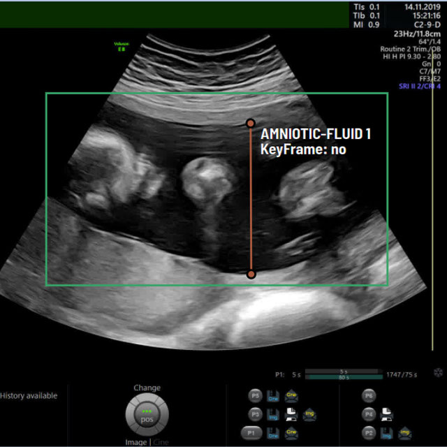
A-AFMA
Challenge UserPrenatal ultrasound (US) measurement of amniotic fluid is an important part of fetal surveillance as it provides a non-invasive way of assessing whether there is oligohydramnios (insufficient amniotic fluid) and polyhydramnios (excess amniotic fluid), which are associated with numerous problems both during pregnancy and after birth. In this Image Analysis Challenge, we aim to attract attention from the image analysis community to work on the problem of automated measurement of the MVP using the predefined ultrasound video clip based on a linear-sweep protocol [1]. We define two tasks. The first task is to automatically detect amniotic fluid and the maternal bladder. The second task is to identify the appropriate points for MVP measurement given the selected frame of the video clip, and calculate the length of the connected line between these points. The data was collected from women in the second trimester of pregnancy, as part of the PURE study at the John Radcliffe Hospital in Oxford, UK.


Breast Cancer Segmentation
Challenge UserSemantic segmentation of histologic regions in scanned FFPE H&E stained slides of triple-negative breast cancer from The Cancer Genome Atlas. See: Amgad M, Elfandy H, ..., Gutman DA, Cooper LAD. Structured crowdsourcing enables convolutional segmentation of histology images. Bioinformatics. 2019. doi: 10.1093/bioinformatics/btz083
fastPET-LD
Challenge UserThe purpose of this challenge is the detection of “hot spots” in fast PET scan, that is locations that have an elevated standard uptake value (SUV) and potential clinical significance. Corresponding CT scans are also provided. The ground truth, common to both datasets, was generated by a nuclear medicine expert. It consists of a 3-D segmentation map of the hot spots as well as a text file containing the position and size of 3D cuboid bounding box for each hot spot.

WSSS4LUAD
Challenge UserThe WSSS4LUAD dataset contains over 10,000 patches of lung adenocarcinoma from whole slide images from Guangdong Provincial People's Hospital and TCGA with image-level annotations. The goal of this challenge is to perform semantic segmentation for differentiating three important types of tissues in the WSIs of lung adenocarcinoma, including cancerous epithelial region, cancerous stroma region and normal region. Paticipants have to use image-level annotations to give pixel-level prediction.


Multi-site, Multi-Domain Airway Tree Modeling (ATM’22)
Challenge UserAirway segmentation is a crucial step for the analysis of pulmonary diseases including asthma, bronchiectasis, and emphysema. The accurate segmentation based on X-Ray computed tomography (CT) enables the quantitative measurements of airway dimensions and wall thickness, which can reveal the abnormality of patients with chronic obstructive pulmonary disease (COPD). Besides, the extraction of patient-specific airway models from CT images is required for navigatiisted surgery.
Shifts Challenge 2022
Challenge UserThe goal of the Shifts Challenge 2022 is to raise awareness among the research community about the problems of distributional shift, robustness, and uncertainty estimation, and to identify new solutions to address them. The competition will consist of two new tracks: White Matter Multiple Sclerosis (MS) lesion segmentation in 3D Magnetic Resonance Imaging (MRI) of the brain and Marine cargo vessel power estimation.