sen yang
ys810137152
- China
- Tencent
- Tencent AI Lab
Statistics
- Member for 5 years, 4 months
- 153 challenge submissions
- 1 algorithms run
Activity Overview

IDRiD
Challenge ParticipantThis challenge evaluates automated techniques for analysis of fundus photographs. We target segmentation of retinal lesions like exudates, microaneurysms, and hemorrhages and detection of the optic disc and fovea. Also, we seek grading of fundus images according to the severity level of DR and DME.
BreastPathQ: Cancer Cellularity Challenge 2019
Challenge ParticipantSPIE-AAPM-NCI BreastPathQ:Cancer Circularity Challenge 2019: Participants will be tasked to develop an automated method for analyzing histology patches extracted from whole slide images and assign a score reflecting cancer cellularity for tumor burden assessment in each.


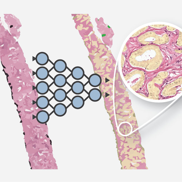
Gleason2019
Challenge ParticipantMICCAI 2019 Automatic Prostate Gleason Grading Challenge: This challenge aims at the automatic Gleason grading of prostate cancer from H&E-stained histopathology images. This task is of critical importance because Gleason score is a strong prognostic predictor. On the other hand, it is very challenging because of the large degree of heterogeneity in the cellular and glandular patterns associated with each Gleason grade, leading to significant inter-observer variability, even among expert pathologists.

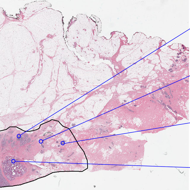
PAIP2020
Challenge ParticipantBuilt on the success of its predecessor, PAIP2020 is the second challenge organized by the Pathology AI Platform (PAIP) and the Seoul National University Hospital (SNUH). PAIP2020 will proceed to not only detect whole tumor areas in colorectal cancers but also to classify their molecular subtypes, which will lead to characterization of their heterogeneity with respect to prognoses and therapeutic responses. All participants should predict one of the molecular carcinogenesis pathways, i.e., microsatellite instability(MSI) in colorectal cancer, by performing digital image analysis without clinical tests. This task has a high clinical relevance as the currently used procedure requires an extensive microscopic assessment by pathologists. Therefore, those automated algorithms would reduce the workload of pathologists as a diagnostic assistance.
Thyroid Nodule Segmentation and Classification
Challenge ParticipantThe main topic of this TN-SCUI2020 challenge is finding automatic algorithms to accurately classify the thyroid nodules in ultrasound images. It will provide the biggest public dataset of thyroid nodule with over 4500 patient cases from different ages, genders, and were collected using different ultrasound machines. Each ultrasound image is provided with its ground truth class (benign or maglinant) and a detailed delineation of the nodule. This challenge will provide a unique opportunity for participants from different backgrounds (e.g. academia, industry, and government, etc.) to compare their algorithms in an impartial way.

CADA
Challenge ParticipantCerebral aneurysms are local dilations of arterial blood vessels caused by a weakness of the vessel wall. Subarachnoid hemorrhage (SAH) caused by the rupture of a cerebral aneurysm is a life-threatening condition associated with high mortality and morbidity. The mortality rate is above 40%, and even in case of survival cognitive impairment can affect patients for a long time. Major goals in image analysis are the detection and risk assessment of aneurysms. We, therefore, subdivided the challenge into three categories. The first task is finding the aneurysm; the second task is the accurate segmentation to allow for a longitudinal assessment of the development of suspicious aneurysms. The third task is the estimation of the rupture risk of the aneurysm.
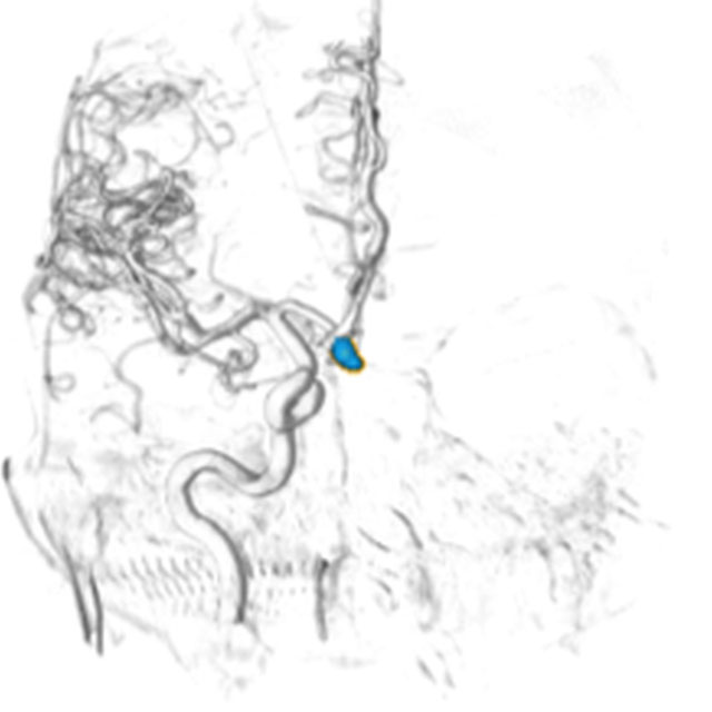
CADA - Aneurysm Segmentation
Challenge ParticipantCerebral aneurysms are local dilations of arterial blood vessels caused by a weakness of the vessel wall. Subarachnoid hemorrhage (SAH) caused by the rupture of a cerebral aneurysm is a life-threatening condition associated with high mortality and morbidity. The mortality rate is above 40%, and even in case of survival cognitive impairment can affect patients for a long time. Major goals in image analysis are the detection and risk assessment of aneurysms. We, therefore, subdivided the challenge into three categories. The first task is finding the aneurysm; the second task is the accurate segmentation to allow for a longitudinal assessment of the development of suspicious aneurysms. The third task is the estimation of the rupture risk of the aneurysm.
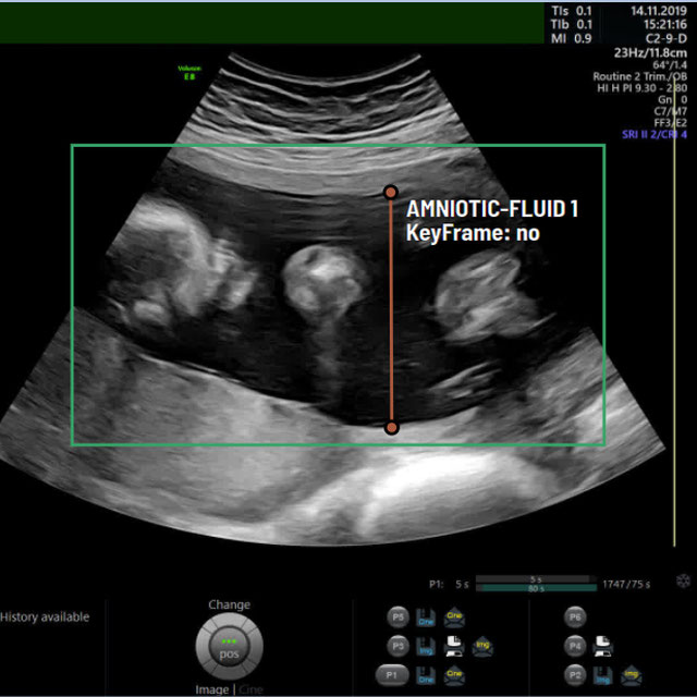
A-AFMA
Challenge ParticipantPrenatal ultrasound (US) measurement of amniotic fluid is an important part of fetal surveillance as it provides a non-invasive way of assessing whether there is oligohydramnios (insufficient amniotic fluid) and polyhydramnios (excess amniotic fluid), which are associated with numerous problems both during pregnancy and after birth. In this Image Analysis Challenge, we aim to attract attention from the image analysis community to work on the problem of automated measurement of the MVP using the predefined ultrasound video clip based on a linear-sweep protocol [1]. We define two tasks. The first task is to automatically detect amniotic fluid and the maternal bladder. The second task is to identify the appropriate points for MVP measurement given the selected frame of the video clip, and calculate the length of the connected line between these points. The data was collected from women in the second trimester of pregnancy, as part of the PURE study at the John Radcliffe Hospital in Oxford, UK.


Breast Cancer Segmentation
Challenge ParticipantSemantic segmentation of histologic regions in scanned FFPE H&E stained slides of triple-negative breast cancer from The Cancer Genome Atlas. See: Amgad M, Elfandy H, ..., Gutman DA, Cooper LAD. Structured crowdsourcing enables convolutional segmentation of histology images. Bioinformatics. 2019. doi: 10.1093/bioinformatics/btz083

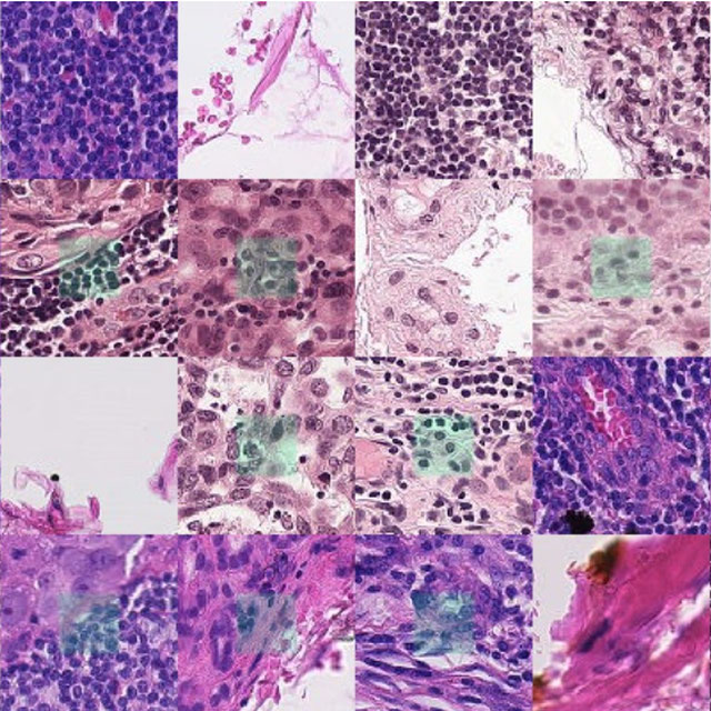
WSSS4LUAD
Challenge ParticipantThe WSSS4LUAD dataset contains over 10,000 patches of lung adenocarcinoma from whole slide images from Guangdong Provincial People's Hospital and TCGA with image-level annotations. The goal of this challenge is to perform semantic segmentation for differentiating three important types of tissues in the WSIs of lung adenocarcinoma, including cancerous epithelial region, cancerous stroma region and normal region. Paticipants have to use image-level annotations to give pixel-level prediction.