Bhavesh Parmar
bhparmar
- India
- L. D. College of Engineering, Gujarat Technological University
- Biomedical Engineering
Statistics
- Member for 5 years, 4 months
- 14 challenge submissions
Activity Overview
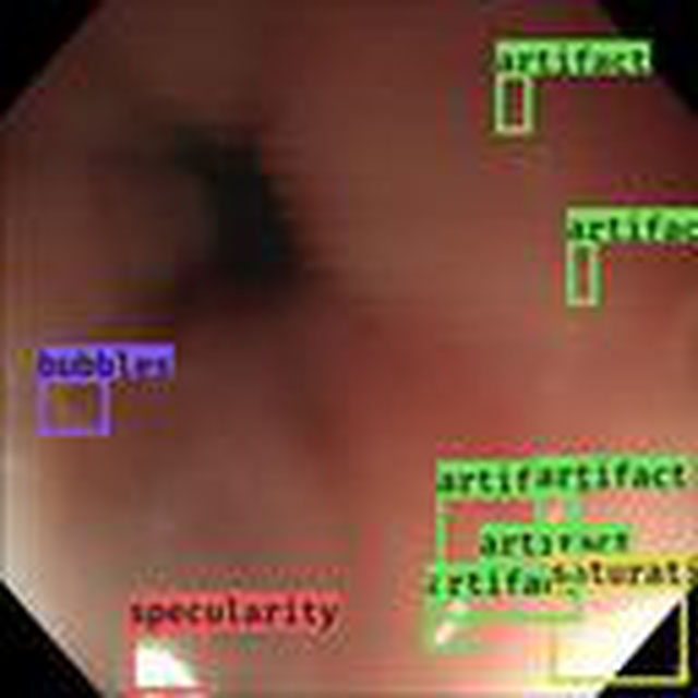


KNOAP2020
Challenge UserKnee osteoarthritis causes a large economic burden on the society and reduces life quality of an individual. Therefore, methods that are able to identify subjects who will develop the disease in the future are important. Usually the methods are optimized for specific datasets and it is unclear how the different methods would perform on previously unseen data. Therefore, we are organizing a challenge to objectively compare methods that use MRI, X-ray image data, and clinical risk factors for prediction of incident symptomatic radiographic knee osteoarthritis.

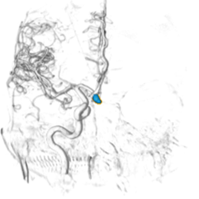
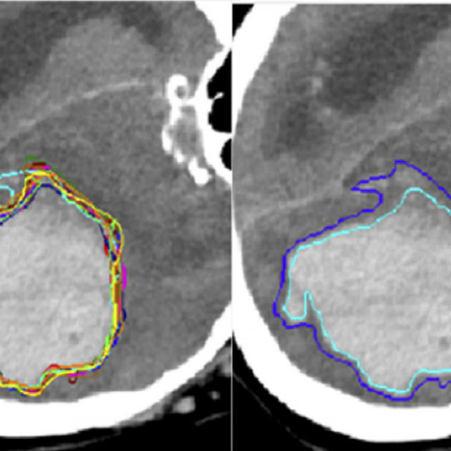
CADA
Challenge UserCerebral aneurysms are local dilations of arterial blood vessels caused by a weakness of the vessel wall. Subarachnoid hemorrhage (SAH) caused by the rupture of a cerebral aneurysm is a life-threatening condition associated with high mortality and morbidity. The mortality rate is above 40%, and even in case of survival cognitive impairment can affect patients for a long time. Major goals in image analysis are the detection and risk assessment of aneurysms. We, therefore, subdivided the challenge into three categories. The first task is finding the aneurysm; the second task is the accurate segmentation to allow for a longitudinal assessment of the development of suspicious aneurysms. The third task is the estimation of the rupture risk of the aneurysm.


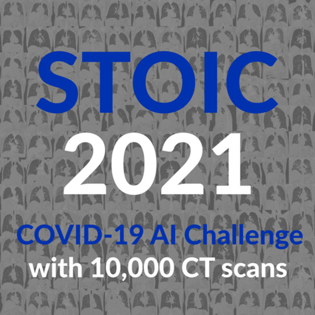
CT diagnosis of COVID-19
Challenge UserCoronavirus disease 2019 (COVID-19) has infected more than 1.3 million individuals all over the world and caused more than 106,000 deaths. One major hurdle in controlling the spreading of this disease is the inefficiency and shortage of medical tests. To mitigate the inefficiency and shortage of existing tests for COVID-19, we propose this competition to encourage the development of effective Deep Learning techniques to diagnose COVID-19 based on CT images. The problem we want to solve is to classify each CT image into positive COVID-19 (the image has clinical findings of COVID-19) or negative COVID-19 ( the image does not have clinical findings of COVID-19). It’s a binary classification problem based on CT images.

CADA - Rupture Risk Estimation
Challenge UserCerebral aneurysms are local dilations of arterial blood vessels caused by a weakness of the vessel wall. Subarachnoid hemorrhage (SAH) caused by the rupture of a cerebral aneurysm is a life-threatening condition associated with high mortality and morbidity. The mortality rate is above 40%, and even in case of survival cognitive impairment can affect patients for a long time. Major goals in image analysis are the detection and risk assessment of aneurysms. We, therefore, subdivided the challenge into three categories. The first task is finding the aneurysm; the second task is the accurate segmentation to allow for a longitudinal assessment of the development of suspicious aneurysms. The third task is the estimation of the rupture risk of the aneurysm.
CADA - Aneurysm Segmentation
Challenge UserCerebral aneurysms are local dilations of arterial blood vessels caused by a weakness of the vessel wall. Subarachnoid hemorrhage (SAH) caused by the rupture of a cerebral aneurysm is a life-threatening condition associated with high mortality and morbidity. The mortality rate is above 40%, and even in case of survival cognitive impairment can affect patients for a long time. Major goals in image analysis are the detection and risk assessment of aneurysms. We, therefore, subdivided the challenge into three categories. The first task is finding the aneurysm; the second task is the accurate segmentation to allow for a longitudinal assessment of the development of suspicious aneurysms. The third task is the estimation of the rupture risk of the aneurysm.

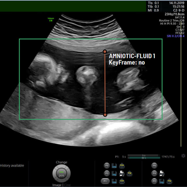
A-AFMA
Challenge UserPrenatal ultrasound (US) measurement of amniotic fluid is an important part of fetal surveillance as it provides a non-invasive way of assessing whether there is oligohydramnios (insufficient amniotic fluid) and polyhydramnios (excess amniotic fluid), which are associated with numerous problems both during pregnancy and after birth. In this Image Analysis Challenge, we aim to attract attention from the image analysis community to work on the problem of automated measurement of the MVP using the predefined ultrasound video clip based on a linear-sweep protocol [1]. We define two tasks. The first task is to automatically detect amniotic fluid and the maternal bladder. The second task is to identify the appropriate points for MVP measurement given the selected frame of the video clip, and calculate the length of the connected line between these points. The data was collected from women in the second trimester of pregnancy, as part of the PURE study at the John Radcliffe Hospital in Oxford, UK.


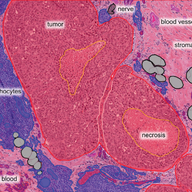
Breast Cancer Segmentation
Challenge UserSemantic segmentation of histologic regions in scanned FFPE H&E stained slides of triple-negative breast cancer from The Cancer Genome Atlas. See: Amgad M, Elfandy H, ..., Gutman DA, Cooper LAD. Structured crowdsourcing enables convolutional segmentation of histology images. Bioinformatics. 2019. doi: 10.1093/bioinformatics/btz083
fastPET-LD
Challenge UserThe purpose of this challenge is the detection of “hot spots” in fast PET scan, that is locations that have an elevated standard uptake value (SUV) and potential clinical significance. Corresponding CT scans are also provided. The ground truth, common to both datasets, was generated by a nuclear medicine expert. It consists of a 3-D segmentation map of the hot spots as well as a text file containing the position and size of 3D cuboid bounding box for each hot spot.
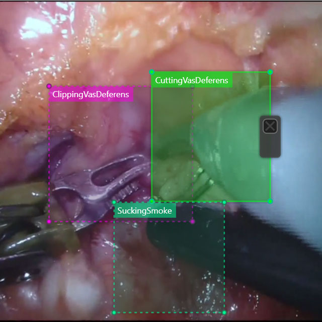
SARAS-MESAD
Challenge UserThis challenge is organized under MICCAI 2021, the 24th International Conference on Medical Image Computing and Computer Assisted Intervention. The event will be held from September 27th to October 1st 2021 in Strasbourg, France. The challenge focuses on multi-domain surgeon action detection.

WSSS4LUAD
Challenge UserThe WSSS4LUAD dataset contains over 10,000 patches of lung adenocarcinoma from whole slide images from Guangdong Provincial People's Hospital and TCGA with image-level annotations. The goal of this challenge is to perform semantic segmentation for differentiating three important types of tissues in the WSIs of lung adenocarcinoma, including cancerous epithelial region, cancerous stroma region and normal region. Paticipants have to use image-level annotations to give pixel-level prediction.

Parse2022
Challenge UserIt is of significant clinical interest to study pulmonary artery structures in the field of medical image analysis. One prerequisite step is to segment pulmonary artery structures from CT with high accuracy and low time-consuming. The segmentation of pulmonary artery structures benefits the quantification of its morphological changes for diagnosis of pulmonary hypertension and thoracic surgery. However, due to the complexity of pulmonary artery topology, automated segmentation of pulmonary artery topology is a challenging task. Besides, the open accessible large-scale CT data with well labeled pulmonary artery are scarce (The large variations of the topological structures from different patients make the annotation an extremely challenging process). The lack of well labeled pulmonary artery hinders the development of automatic pulmonary artery segmentation algorithm. Hence, we try to host the first Pulmonary ARtery SEgmentation challenge in MICCAI 2022 (Named Parse2022) to start a new research topic.
DFUC 2022
Challenge UserDiabetes is a global epidemic affecting around 425 million people and expected to rise to 629 million by 2045. Diabetic Foot Ulcer (DFU) is a severe condition that can result from the disease. The rise of the condition over the last decades is a challenge for healthcare systems. Cases of DFU usually lead to severe conditions that greatly prolongs treatment and result in limb amputation or death. Recent research focuses on creating detection algorithms to monitor their condition to improve patient care and reduce strain on healthcare systems. Work between Manchester Metropolitan University, Lancashire Teaching Hospitals and Manchester University NHS Foundation Trust has created an international repository of up to 11,000 DFU images. Analysis of ulcer regions is a key for DFU management. Delineation of ulcers is time-consuming. With effort from the lead scientists of the UK, US, India and New Zealand, this challenge promotes novel work in DFU segmentation and promote interdisciplinary researcher collaboration.
3D Teeth Scan Segmentation and Labeling Challenge MICCAI2022
Challenge UserComputer-aided design (CAD) tools have become increasingly popular in modern dentistry for highly accurate treatment planning. In particular, in orthodontic CAD systems, advanced intraoral scanners (IOSs) are now widely used as they provide precise digital surface models of the dentition. Such models can dramatically help dentists simulate teeth extraction, move, deletion, and rearrangement and therefore ease the prediction of treatment outcomes. Although IOSs are becoming widespread in clinical dental practice, there are only few contributions on teeth segmentation/labeling available in the literature and no publicly available database. A fundamental issue that appears with IOS data is the ability to reliably segment and identify teeth in scanned observations. Teeth segmentation and labelling is difficult as a result of the inherent similarities between teeth shapes as well as their ambiguous positions on jaws.


Multi-site, Multi-Domain Airway Tree Modeling (ATM’22)
Challenge UserAirway segmentation is a crucial step for the analysis of pulmonary diseases including asthma, bronchiectasis, and emphysema. The accurate segmentation based on X-Ray computed tomography (CT) enables the quantitative measurements of airway dimensions and wall thickness, which can reveal the abnormality of patients with chronic obstructive pulmonary disease (COPD). Besides, the extraction of patient-specific airway models from CT images is required for navigatiisted surgery.


SEG.A. - Segmentation of the Aorta
Challenge UserSegmentation, modeling and visualization of the arterial tree are still a challenge in medical image analysis. The main track of this challenge deals with the fully automatic segmentation of the aortic vessel tree in computed tomography images. Optionally, teams can submit tailored solutions for meshing and visualization of the vessel tree.


Light My Cells : Bright Field to Fluorescence Imaging Challenge
Challenge UserJoin the Light My Cells France-Bioimaging challenge! Enhance biology and microscopy by contributing to the development of new image-to-image deep labelling methods. The task: predict the best-focused output images of several fluorescently labelled organelles from label-free transmitted light input images. Dive into the future of imaging with us! 🌐🔬 #LightMyCellsChallenge
Diminished Reality for Emerging Applications in Medicine
Challenge UserThe Diminished Reality for Emerging Applications in Medicine through Inpainting (DREAMING) challenge seeks to pioneer the integration of Diminished Reality (DR) into oral and maxillofacial surgery. While Augmented Reality (AR) has been extensively explored in medicine, DR remains largely uncharted territory. DR involves virtually removing real objects from the environment by replacing them with their background. Recent inpainting methods present an opportunity for real-time DR applications without scene knowledge. DREAMING focuses on implementing such methods to fill obscured regions in surgery scenes with realistic backgrounds, emphasizing the complex facial anatomy and patient diversity. The challenge provides a dataset of synthetic yet photorealistic surgery scenes featuring humans, simulating an operating room setting. Participants are tasked with developing algorithms that seamlessly remove disruptions caused by medical instruments and hands, offering surgeons an unimpeded view of the operative site.