Your mugshot

Willam Green

dskswu

  •  United States of America
  •  Student
  •  Student
Statistics
  • Member for 5 years, 2 months

Activity Overview

ICIAR2018-Challenge Logo
ICIAR 2018
Challenge User

Can you develop a method for automatic detection of cancerous regions in breast cancer histology images?

CHAOS Logo
CHAOS
Challenge User

In this challenge, you segment the liver in CT data, and segment liver, spleen, and kidneys in MRI data.

ACDC-LungHP Logo
ACDC-LungHP
Challenge User

Automatic Cancer Detection and Classification in Whole-slide Lung Histopathology

Decathlon-10 Logo
Decathlon
Challenge User

The Medical Segmentation Decathlon challenge tests the generalisability of machine learning algorithms when applied to 10 different semantic segmentation task.

KiTS19 Logo
KiTS19
Challenge User

2019 Kidney and Kidney Tumor Segmentation Challenge

PAIP2019 Logo
PAIP 2019
Challenge User

PAIP2019: Liver Cancer Segmentation Task 1: Liver Cancer Segmentation Task 2: Viable Tumor Burden Estimation

curious2019 Logo
curious2019
Challenge User

MICCAI Challenge 2019 for Correction of Brainshift with Intra-Operative Ultrasound. Taks 1: Register pre-operative MRI to iUS before tumor resection;Taks 2: Register iUS after tumor resection to iUS before tumor resection

VerSe2019 Logo
VerSe`19
Challenge User

Vertebrae labelling and segmentation on a spine dataset on an unprecedented 150 CT scans with voxel-level vertebral annotations.

LYSTO Logo
Lymphocyte Assessment Hackathon
Challenge User

Lymphocyte Assessment Hackathon in conjunction with the MICCAI COMPAY 2019 Workshop on Computational Pathology

ECDP2020 Logo
HEROHE
Challenge User

Unlike previous challenges, this proposes to find an image analysis algorithm to identify HER2-positive from HER2-negative breast cancer specimens evaluating only the morphological features present on the HE slide, without the staining patterns of IHC.

EndoCV Logo
EndoCV2020
Challenge User

Endoscopy computer vision challenge (EndoCV2020) introduces two core sub-themes in endoscopy: 1) artefact detection and segmentation (EAD2020) and 2) disease detection and segmentation (EDD2020).

LNDb Logo
LNDb Challenge
Challenge User

Lung cancer screening and Fleischner follow-up determination in chest CT through nodule detection, segmentation and characterization

PAIP2020 Logo
PAIP2020
Challenge User

Built on the success of its predecessor, PAIP2020 is the second challenge organized by the Pathology AI Platform (PAIP) and the Seoul National University Hospital (SNUH). PAIP2020 will proceed to not only detect whole tumor areas in colorectal cancers but also to classify their molecular subtypes, which will lead to characterization of their heterogeneity with respect to prognoses and therapeutic responses. All participants should predict one of the molecular carcinogenesis pathways, i.e., microsatellite instability(MSI) in colorectal cancer, by performing digital image analysis without clinical tests. This task has a high clinical relevance as the currently used procedure requires an extensive microscopic assessment by pathologists. Therefore, those automated algorithms would reduce the workload of pathologists as a diagnostic assistance.

RibFrac Logo
RibFrac
Challenge User

Rib Fracture Detection and Classification Challenge: A large-scale benchmark of 660 CT scans with ~5,000 rib fractures (around 80Gb)

TN-SCUI2020 Logo
Thyroid Nodule Segmentation and Classification
Challenge User

The main topic of this TN-SCUI2020 challenge is finding automatic algorithms to accurately classify the thyroid nodules in ultrasound images. It will provide the biggest public dataset of thyroid nodule with over 4500 patient cases from different ages, genders, and were collected using different ultrasound machines. Each ultrasound image is provided with its ground truth class (benign or maglinant) and a detailed delineation of the nodule. This challenge will provide a unique opportunity for participants from different backgrounds (e.g. academia, industry, and government, etc.) to compare their algorithms in an impartial way.

SARAS-ESAD
Challenge User

This challenge is part of Medical Imaging with Deep Learning conference, 2020. The conference is held between 6 ‑ 8 July 2020 in Montréal. The SARAS (Smart Autonomous Robotic Assistant Surgeon) EU consortium, www.saras-project.eu, is working towards replacing the assistant surgeon in MIS with two assistive robotic arms. To accomplish that, an artificial intelligence based system is required which not only can understand the complete surgical scene but also detect the actions being performed by the main surgeon. This information can later be used infer the response required from the autonomous assistant surgeon.

DFU2020 Logo
Diabetic Foot Ulcer Challenge 2020
Challenge User

Diabetic Foot Ulcer Challenge 2020

COVID-CT Logo
CT diagnosis of COVID-19
Challenge User

Coronavirus disease 2019 (COVID-19) has infected more than 1.3 million individuals all over the world and caused more than 106,000 deaths. One major hurdle in controlling the spreading of this disease is the inefficiency and shortage of medical tests. To mitigate the inefficiency and shortage of existing tests for COVID-19, we propose this competition to encourage the development of effective Deep Learning techniques to diagnose COVID-19 based on CT images. The problem we want to solve is to classify each CT image into positive COVID-19 (the image has clinical findings of COVID-19) or negative COVID-19 ( the image does not have clinical findings of COVID-19). It’s a binary classification problem based on CT images.
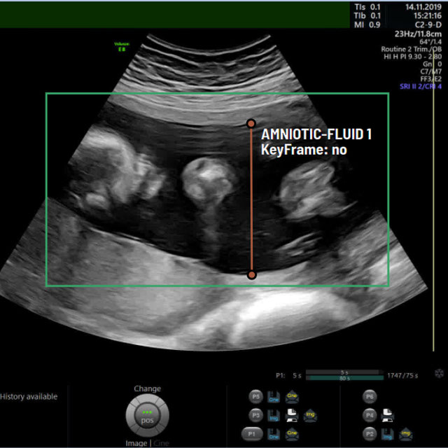
lodopab Logo
LoDoPaB-CT
Challenge User

Low-Dose CT reconstruction in the setting of the LoDoPaB-CT dataset.

RIADD Logo
RIADD (ISBI-2021)
Challenge User

Retinal Image Analysis for multi-Disease Detection

MitoEM Logo
MitoEM
Challenge User

Large-scale 3D mitochondria instance segmentation benchmark

A-AFMA Logo
A-AFMA
Challenge User

Prenatal ultrasound (US) measurement of amniotic fluid is an important part of fetal surveillance as it provides a non-invasive way of assessing whether there is oligohydramnios (insufficient amniotic fluid) and polyhydramnios (excess amniotic fluid), which are associated with numerous problems both during pregnancy and after birth. In this Image Analysis Challenge, we aim to attract attention from the image analysis community to work on the problem of automated measurement of the MVP using the predefined ultrasound video clip based on a linear-sweep protocol [1]. We define two tasks. The first task is to automatically detect amniotic fluid and the maternal bladder. The second task is to identify the appropriate points for MVP measurement given the selected frame of the video clip, and calculate the length of the connected line between these points. The data was collected from women in the second trimester of pregnancy, as part of the PURE study at the John Radcliffe Hospital in Oxford, UK.

covid-segmentation Logo
COVID-19 LUNG CT LESION SEGMENTATION CHALLENGE - 2020
Challenge User

This challenge will create the platform to evaluate emerging methods for the segmentation and quantification of lung lesions caused by SARS-CoV-2 infection from CT images.

VALDO Logo
Where is VALDO?
Challenge User

Vascular Lesion Detection Challenge at MICCAI 2021

SegPC-2021 Logo
SegPC-2021
Challenge User

This challenge is positioned towards robust segmentation of cells which is the first stage to build such a tool for plasma cell cancer, namely, Multiple Myeloma (MM), which is a type of blood cancer.

FUSC Logo
Foot Ulcer Segmentation Challenge
Challenge User

Carotid Artery Vessel Wall Segmentation Challenge
Challenge User

To segment the vessel wall of the carotid artery on black-blood MRI images

crossMoDA Logo
Cross-Modality Domain Adaptation Image Segmentation - 2021
Challenge User

The CrossMoDA challenge 2021 introduces the first large and multi-class medical dataset for unsupervised cross-modality Domain Adaptation.

kits21 Logo
KiTS21
Challenge User

The 2021 MICCAI Kidney and Kidney Tumor Segmentation challenge

CholecTriplet2021 Logo
CholecTriplet 2021
Challenge User

EndoVis Sub-challenge for Surgical Action Triplet Recognition

PAIP2021 Logo
PAIP2021
Challenge User

PAIP 2021 Challenge; Perineural invasion in multiple organ cancer (colon, prostate and pancreatobiliary tract)

NuCLS Logo
NuCLS
Challenge User

Classification, Localization and Segmentation of nuclei in scanned FFPE H&E stained slides of triple-negative breast cancer from The Cancer Genome Atlas. See: Amgad et al. 2021. arXiv:2102.09099 [cs.CV].

BCSegmentation Logo
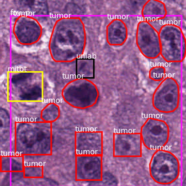
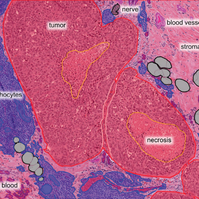
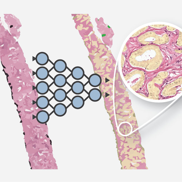
Breast Cancer Segmentation
Challenge User

Semantic segmentation of histologic regions in scanned FFPE H&E stained slides of triple-negative breast cancer from The Cancer Genome Atlas. See: Amgad M, Elfandy H, ..., Gutman DA, Cooper LAD. Structured crowdsourcing enables convolutional segmentation of histology images. Bioinformatics. 2019. doi: 10.1093/bioinformatics/btz083

feta Logo
FeTA - Fetal Tissue Annotation Challenge
Challenge User

Fetal Tissue Annotation Challenge

DFU-2021 Logo
DFUC2021
Challenge User

Diabetic Foot Ulcer Challenge 2021

NODE21 Logo
NODE21
Challenge User

NODE21: generate and detect nodules on chest radiographs

WSSS4LUAD Logo
WSSS4LUAD
Challenge User

The WSSS4LUAD dataset contains over 10,000 patches of lung adenocarcinoma from whole slide images from Guangdong Provincial People's Hospital and TCGA with image-level annotations. The goal of this challenge is to perform semantic segmentation for differentiating three important types of tissues in the WSIs of lung adenocarcinoma, including cancerous epithelial region, cancerous stroma region and normal region. Paticipants have to use image-level annotations to give pixel-level prediction.

CXR-COVID19 Logo
Chest XR COVID-19 detection
Challenge User

Build AI models to detect COVID-19 using Chest X-ray images

PI-CAI Logo
The PI-CAI Challenge
Challenge User

Artificial Intelligence and Radiologists at Prostate Cancer Detection in MRI

CoNIC-Challenge Logo
CoNIC 2022
Challenge User

Colon Nuclei Identification and Counting Challenge 2022

BCNB Logo
BCNB
Challenge User

Early Breast Cancer Core-Needle Biopsy WSI Dataset

autoPET Logo
autoPET
Challenge User

Automatic lesion segmentation in whole-body FDG-PET/CT

DRAC22 Logo
Diabetic Retinopathy Analysis Challenge MICCAI2022
Challenge User

Diabetic Retinopathy (DR) lesions segmentation, image quality assessment and classification of proliferatived DR (PDR) and non-PDR in ultra-wide optical coherence tomography angiography mosaic (UW-OCTA-M) images

vessel-wall-segmentation-2022 Logo
Carotid Vessel Wall Segmentation and Atherosclerosis Diagnosis
Challenge User

ATM22 Logo
Multi-site, Multi-Domain Airway Tree Modeling (ATM’22)
Challenge User

Airway segmentation is a crucial step for the analysis of pulmonary diseases including asthma, bronchiectasis, and emphysema. The accurate segmentation based on X-Ray computed tomography (CT) enables the quantitative measurements of airway dimensions and wall thickness, which can reveal the abnormality of patients with chronic obstructive pulmonary disease (COPD). Besides, the extraction of patient-specific airway models from CT images is required for navigatiisted surgery.

isles22 Logo
Ischemic Stroke Lesion Segmentation Challenge
Challenge User

bci Logo
Breast Cancer Immunohistochemical Image Generation Challenge
Challenge User

The Breast Cancer Immunohistochemical Image Generation Challenge aims to directly generate IHC-stained breast cancer histopathology images from HE-stained images.

2023PAIP Logo
PAIP 2023: TC prediction in pancreatic and colon cancer
Challenge User

Tumor cellularity prediction in pancreatic cancer (supervised learning) and colon cancer (transfer learning)

toothfairy Logo
ToothFairy: Cone-Beam Computed Tomography Segmentation Challenge
Challenge User

This is the first edition of the ToothFairy challenge organized by the University of Modena and Reggio Emilia with the collaboration of Raudboud University. This challenge aims at pushing the development of deep learning frameworks to segment the Inferior Alveolar Canal (IAC) by incrementally extending the amount of publicly available 3D-annotated Cone Beam Computed Tomography (CBCT) scans. CBCT modality is becoming increasingly important for treatment planning and diagnosis in implant dentistry and maxillofacial surgery. The three-dimensional information acquired with CBCT can be crucial to plan a vast number of surgical interventions with the aim of preserving noble anatomical structures such as the Inferior Alveolar Canal (IAC), which contains the homonymous nerve (Inferior Alveolar Nerve, IAN). Deep learning models can support medical personnel in surgical planning procedures by providing a voxel-level segmentation of the IAN automatically extracted from CBCT scans.

ARCADE Logo
ARCADE-MICCAI2023
Challenge User

MedFM2023 Logo
Foundation Model Prompting for Medical Image Classification
Challenge User

The primary objective of this challenge is to promote the development and evaluation of model adaptation techniques for medical image classification to leverage the existing foundation models.

DENTEX Logo
DENTEX - MICCAI23
Challenge User

Dental Enumeration and Diagnosis on Panoramic X- rays Challenge

THOMPSON-Challenge Logo
The Trauma THOMPSON Challenge
Challenge User

JustRAIGS Logo
Justified Referral in AI Glaucoma Screening
Challenge User

LEOPARD Logo
The LEOPARD Challenge
Challenge User

Gleason Grading of Prostate Biopsies Logo
Gleason Grading of Prostate Biopsies
Algorithm User

Automated Gleason grading of prostate biopsies following the Gleason Grade Group system.