Albert Yee
Yeah
- China
- Southern University of Science and Technology
- iMED
- Website
Statistics
- Member for 5 years, 1 month
- 83 challenge submissions
Activity Overview
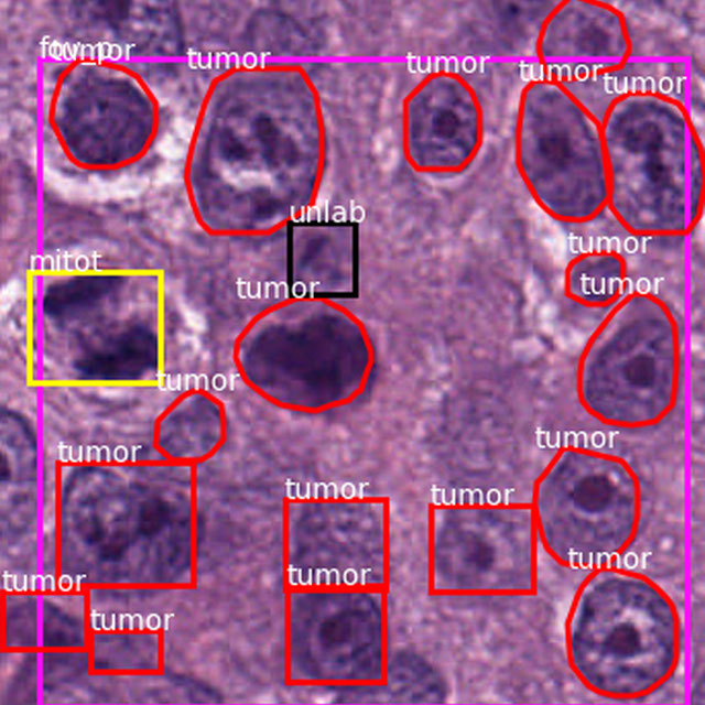
BreastPathQ: Cancer Cellularity Challenge 2019
Challenge UserSPIE-AAPM-NCI BreastPathQ:Cancer Circularity Challenge 2019: Participants will be tasked to develop an automated method for analyzing histology patches extracted from whole slide images and assign a score reflecting cancer cellularity for tumor burden assessment in each.


PAIP2020
Challenge UserBuilt on the success of its predecessor, PAIP2020 is the second challenge organized by the Pathology AI Platform (PAIP) and the Seoul National University Hospital (SNUH). PAIP2020 will proceed to not only detect whole tumor areas in colorectal cancers but also to classify their molecular subtypes, which will lead to characterization of their heterogeneity with respect to prognoses and therapeutic responses. All participants should predict one of the molecular carcinogenesis pathways, i.e., microsatellite instability(MSI) in colorectal cancer, by performing digital image analysis without clinical tests. This task has a high clinical relevance as the currently used procedure requires an extensive microscopic assessment by pathologists. Therefore, those automated algorithms would reduce the workload of pathologists as a diagnostic assistance.
SARAS-ESAD
Challenge UserThis challenge is part of Medical Imaging with Deep Learning conference, 2020. The conference is held between 6 ‑ 8 July 2020 in Montréal. The SARAS (Smart Autonomous Robotic Assistant Surgeon) EU consortium, www.saras-project.eu, is working towards replacing the assistant surgeon in MIS with two assistive robotic arms. To accomplish that, an artificial intelligence based system is required which not only can understand the complete surgical scene but also detect the actions being performed by the main surgeon. This information can later be used infer the response required from the autonomous assistant surgeon.
CT diagnosis of COVID-19
Challenge UserCoronavirus disease 2019 (COVID-19) has infected more than 1.3 million individuals all over the world and caused more than 106,000 deaths. One major hurdle in controlling the spreading of this disease is the inefficiency and shortage of medical tests. To mitigate the inefficiency and shortage of existing tests for COVID-19, we propose this competition to encourage the development of effective Deep Learning techniques to diagnose COVID-19 based on CT images. The problem we want to solve is to classify each CT image into positive COVID-19 (the image has clinical findings of COVID-19) or negative COVID-19 ( the image does not have clinical findings of COVID-19). It’s a binary classification problem based on CT images.


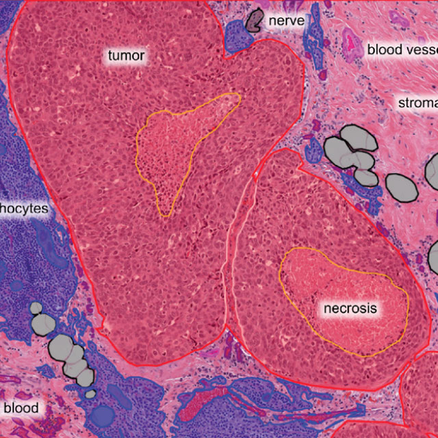
Breast Cancer Segmentation
Challenge UserSemantic segmentation of histologic regions in scanned FFPE H&E stained slides of triple-negative breast cancer from The Cancer Genome Atlas. See: Amgad M, Elfandy H, ..., Gutman DA, Cooper LAD. Structured crowdsourcing enables convolutional segmentation of histology images. Bioinformatics. 2019. doi: 10.1093/bioinformatics/btz083

WSSS4LUAD
Challenge UserThe WSSS4LUAD dataset contains over 10,000 patches of lung adenocarcinoma from whole slide images from Guangdong Provincial People's Hospital and TCGA with image-level annotations. The goal of this challenge is to perform semantic segmentation for differentiating three important types of tissues in the WSIs of lung adenocarcinoma, including cancerous epithelial region, cancerous stroma region and normal region. Paticipants have to use image-level annotations to give pixel-level prediction.
Parse2022
Challenge UserIt is of significant clinical interest to study pulmonary artery structures in the field of medical image analysis. One prerequisite step is to segment pulmonary artery structures from CT with high accuracy and low time-consuming. The segmentation of pulmonary artery structures benefits the quantification of its morphological changes for diagnosis of pulmonary hypertension and thoracic surgery. However, due to the complexity of pulmonary artery topology, automated segmentation of pulmonary artery topology is a challenging task. Besides, the open accessible large-scale CT data with well labeled pulmonary artery are scarce (The large variations of the topological structures from different patients make the annotation an extremely challenging process). The lack of well labeled pulmonary artery hinders the development of automatic pulmonary artery segmentation algorithm. Hence, we try to host the first Pulmonary ARtery SEgmentation challenge in MICCAI 2022 (Named Parse2022) to start a new research topic.
Multi-site, Multi-Domain Airway Tree Modeling (ATM’22)
Challenge UserAirway segmentation is a crucial step for the analysis of pulmonary diseases including asthma, bronchiectasis, and emphysema. The accurate segmentation based on X-Ray computed tomography (CT) enables the quantitative measurements of airway dimensions and wall thickness, which can reveal the abnormality of patients with chronic obstructive pulmonary disease (COPD). Besides, the extraction of patient-specific airway models from CT images is required for navigatiisted surgery.
Shifts Challenge 2022
Challenge UserThe goal of the Shifts Challenge 2022 is to raise awareness among the research community about the problems of distributional shift, robustness, and uncertainty estimation, and to identify new solutions to address them. The competition will consist of two new tracks: White Matter Multiple Sclerosis (MS) lesion segmentation in 3D Magnetic Resonance Imaging (MRI) of the brain and Marine cargo vessel power estimation.
SEG.A. - Segmentation of the Aorta
Challenge UserSegmentation, modeling and visualization of the arterial tree are still a challenge in medical image analysis. The main track of this challenge deals with the fully automatic segmentation of the aortic vessel tree in computed tomography images. Optionally, teams can submit tailored solutions for meshing and visualization of the vessel tree.