Your mugshot

Shuoyu Xu

Biototem

  •  China
  •  Bio-totem Pte Ltd
  •  Digital Pathology
Statistics
  • Member for 4 years, 8 months
  • 137 challenge submissions
  • 82 algorithms run

Activity Overview

CAMELYON17 Logo
CAMELYON17
Challenge User

Automated detection and classification of breast cancer metastases in whole-slide images of histological lymph node sections. This task has high clinical relevance and would normally require extensive microscopic assessment by pathologists.

ICIAR2018-Challenge Logo
ICIAR 2018
Challenge User

Can you develop a method for automatic detection of cancerous regions in breast cancer histology images?

BreastPathQ Logo
BreastPathQ: Cancer Cellularity Challenge 2019
Challenge User

SPIE-AAPM-NCI BreastPathQ:Cancer Circularity Challenge 2019: Participants will be tasked to develop an automated method for analyzing histology patches extracted from whole slide images and assign a score reflecting cancer cellularity for tumor burden assessment in each.
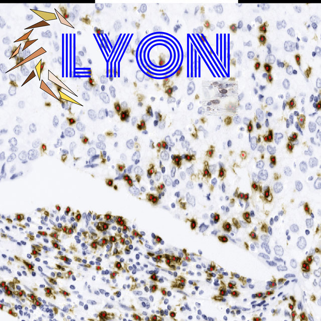
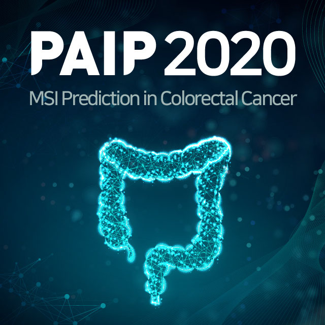
ACDC-LungHP Logo
ACDC-LungHP
Challenge User

Automatic Cancer Detection and Classification in Whole-slide Lung Histopathology

LYON19 Logo
LYON19
Challenge User

Automatic Lymphocyte detection in IHC stained specimens.

PAIP2019 Logo
PAIP 2019
Challenge User

PAIP2019: Liver Cancer Segmentation Task 1: Liver Cancer Segmentation Task 2: Viable Tumor Burden Estimation

LYSTO Logo
Lymphocyte Assessment Hackathon
Challenge User

Lymphocyte Assessment Hackathon in conjunction with the MICCAI COMPAY 2019 Workshop on Computational Pathology

ECDP2020 Logo
HEROHE
Challenge User

Unlike previous challenges, this proposes to find an image analysis algorithm to identify HER2-positive from HER2-negative breast cancer specimens evaluating only the morphological features present on the HE slide, without the staining patterns of IHC.

PAIP2020 Logo
PAIP2020
Challenge User

Built on the success of its predecessor, PAIP2020 is the second challenge organized by the Pathology AI Platform (PAIP) and the Seoul National University Hospital (SNUH). PAIP2020 will proceed to not only detect whole tumor areas in colorectal cancers but also to classify their molecular subtypes, which will lead to characterization of their heterogeneity with respect to prognoses and therapeutic responses. All participants should predict one of the molecular carcinogenesis pathways, i.e., microsatellite instability(MSI) in colorectal cancer, by performing digital image analysis without clinical tests. This task has a high clinical relevance as the currently used procedure requires an extensive microscopic assessment by pathologists. Therefore, those automated algorithms would reduce the workload of pathologists as a diagnostic assistance.

MitoEM Logo
MitoEM
Challenge User

Large-scale 3D mitochondria instance segmentation benchmark

SegPC-2021 Logo
SegPC-2021
Challenge User

This challenge is positioned towards robust segmentation of cells which is the first stage to build such a tool for plasma cell cancer, namely, Multiple Myeloma (MM), which is a type of blood cancer.

PAIP2021 Logo
PAIP2021
Challenge User

PAIP 2021 Challenge; Perineural invasion in multiple organ cancer (colon, prostate and pancreatobiliary tract)

NuCLS Logo
NuCLS
Challenge User

Classification, Localization and Segmentation of nuclei in scanned FFPE H&E stained slides of triple-negative breast cancer from The Cancer Genome Atlas. See: Amgad et al. 2021. arXiv:2102.09099 [cs.CV].

BCSegmentation Logo
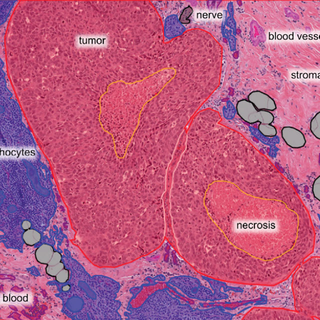
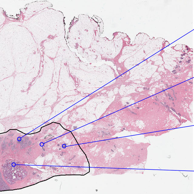
Breast Cancer Segmentation
Challenge User

Semantic segmentation of histologic regions in scanned FFPE H&E stained slides of triple-negative breast cancer from The Cancer Genome Atlas. See: Amgad M, Elfandy H, ..., Gutman DA, Cooper LAD. Structured crowdsourcing enables convolutional segmentation of histology images. Bioinformatics. 2019. doi: 10.1093/bioinformatics/btz083

DFU-2021 Logo
DFUC2021
Challenge User

Diabetic Foot Ulcer Challenge 2021

WSSS4LUAD Logo
WSSS4LUAD
Challenge User

The WSSS4LUAD dataset contains over 10,000 patches of lung adenocarcinoma from whole slide images from Guangdong Provincial People's Hospital and TCGA with image-level annotations. The goal of this challenge is to perform semantic segmentation for differentiating three important types of tissues in the WSIs of lung adenocarcinoma, including cancerous epithelial region, cancerous stroma region and normal region. Paticipants have to use image-level annotations to give pixel-level prediction.

MIDOG2021 Logo
MIDOG Challenge 2021
Challenge User

Mitosis Domain Generalization Challenge 2021 (part of MICCAI 2021)

tiger Logo
TIGER
Challenge User

Grand challenge on automate assessment of tumor infiltrating lymphocytes in digital pathology slides of triple negative and Her2-positive breast cancers

CoNIC-Challenge Logo
CoNIC 2022
Challenge User

Colon Nuclei Identification and Counting Challenge 2022

BCNB Logo
BCNB
Challenge User

Early Breast Cancer Core-Needle Biopsy WSI Dataset

AGGC22 Logo
AGGC22
Challenge User

ACROBAT Logo
ACROBAT 2023
Challenge User

The ACROBAT challenge aims to advance the development of WSI registration algorithms that can align WSIs of IHC-stained breast cancer tissue sections to corresponding tissue regions that were stained with H&E. All WSIs originate from routine diagnostic workflows.

MIDOG2022 Logo
MItosis DOmain Generalization Challenge 2022
Challenge User

NeurIPS22-CellSeg Logo
Cell Segmentation in Multi-modality Microscopy Images
Challenge User

Weakly Supervised Cell Segmentation in Multi-modality High-resolution Microscopy Images

bci Logo
Breast Cancer Immunohistochemical Image Generation Challenge
Challenge User

The Breast Cancer Immunohistochemical Image Generation Challenge aims to directly generate IHC-stained breast cancer histopathology images from HE-stained images.

2023PAIP Logo
PAIP 2023: TC prediction in pancreatic and colon cancer
Challenge User

Tumor cellularity prediction in pancreatic cancer (supervised learning) and colon cancer (transfer learning)

ENDO-AID Logo
Endometrial Carcinoma Detection in Pipelle biopsies
Challenge User

Evaluation platform as reference benchmark for algorithms that can predict endometrial carcinoma on whole-slide images of Pipelle sampled endometrial slides stained in H&E, based on the test data set used in our project.

MedFM2023 Logo
Foundation Model Prompting for Medical Image Classification
Challenge User

The primary objective of this challenge is to promote the development and evaluation of model adaptation techniques for medical image classification to leverage the existing foundation models.

OCELOT2023 Logo
OCELOT 2023: Cell Detection from Cell-Tissue Interaction
Challenge User

lightmycells Logo
Light My Cells : Bright Field to Fluorescence Imaging Challenge
Challenge User

Join the Light My Cells France-Bioimaging challenge! Enhance biology and microscopy by contributing to the development of new image-to-image deep labelling methods. The task: predict the best-focused output images of several fluorescently labelled organelles from label-free transmitted light input images. Dive into the future of imaging with us! 🌐🔬 #LightMyCellsChallenge

LEOPARD Logo
The LEOPARD Challenge
Challenge User

AI4Life-MDC24 Logo
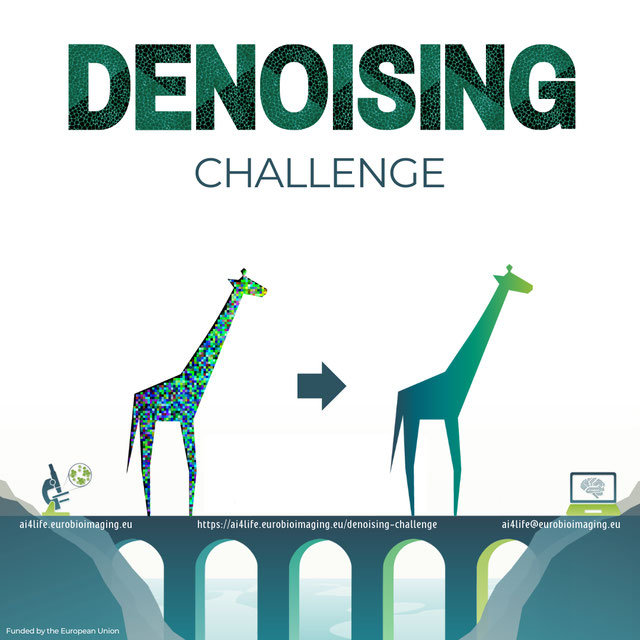
AI4Life Microscopy Denoising Challenge
Challenge User

Wellcome to AI4Life-MDC24! In this challenge, we want to focus on an unsupervised denoising of microscopy images. By participating, researchers can contribute to a critical area of scientific research, aiding in interpreting microscopy images and potentially unlocking discoveries in biology and medicine.