
Yan Zeng
dearxiaojinmao
- China
- Beijing University of Technology
- Department of Biomedical Engineering
Statistics
- Member for 4 years, 7 months
- 87 challenge submissions
Activity Overview
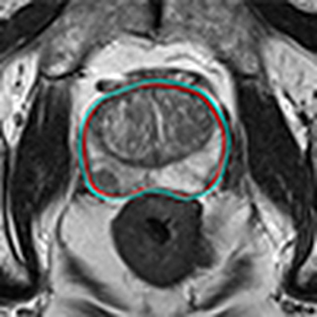

Thyroid Nodule Segmentation and Classification
Challenge UserThe main topic of this TN-SCUI2020 challenge is finding automatic algorithms to accurately classify the thyroid nodules in ultrasound images. It will provide the biggest public dataset of thyroid nodule with over 4500 patient cases from different ages, genders, and were collected using different ultrasound machines. Each ultrasound image is provided with its ground truth class (benign or maglinant) and a detailed delineation of the nodule. This challenge will provide a unique opportunity for participants from different backgrounds (e.g. academia, industry, and government, etc.) to compare their algorithms in an impartial way.
CT diagnosis of COVID-19
Challenge UserCoronavirus disease 2019 (COVID-19) has infected more than 1.3 million individuals all over the world and caused more than 106,000 deaths. One major hurdle in controlling the spreading of this disease is the inefficiency and shortage of medical tests. To mitigate the inefficiency and shortage of existing tests for COVID-19, we propose this competition to encourage the development of effective Deep Learning techniques to diagnose COVID-19 based on CT images. The problem we want to solve is to classify each CT image into positive COVID-19 (the image has clinical findings of COVID-19) or negative COVID-19 ( the image does not have clinical findings of COVID-19). It’s a binary classification problem based on CT images.
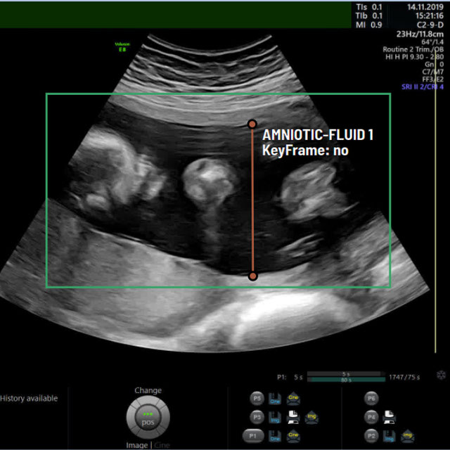
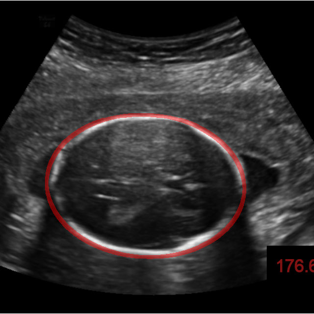
A-AFMA
Challenge UserPrenatal ultrasound (US) measurement of amniotic fluid is an important part of fetal surveillance as it provides a non-invasive way of assessing whether there is oligohydramnios (insufficient amniotic fluid) and polyhydramnios (excess amniotic fluid), which are associated with numerous problems both during pregnancy and after birth. In this Image Analysis Challenge, we aim to attract attention from the image analysis community to work on the problem of automated measurement of the MVP using the predefined ultrasound video clip based on a linear-sweep protocol [1]. We define two tasks. The first task is to automatically detect amniotic fluid and the maternal bladder. The second task is to identify the appropriate points for MVP measurement given the selected frame of the video clip, and calculate the length of the connected line between these points. The data was collected from women in the second trimester of pregnancy, as part of the PURE study at the John Radcliffe Hospital in Oxford, UK.

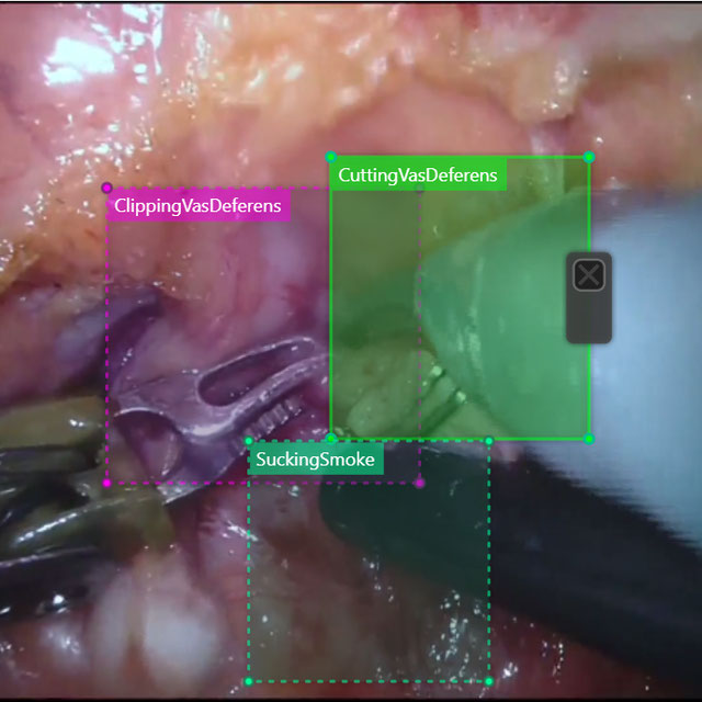
SARAS-MESAD
Challenge UserThis challenge is organized under MICCAI 2021, the 24th International Conference on Medical Image Computing and Computer Assisted Intervention. The event will be held from September 27th to October 1st 2021 in Strasbourg, France. The challenge focuses on multi-domain surgeon action detection.
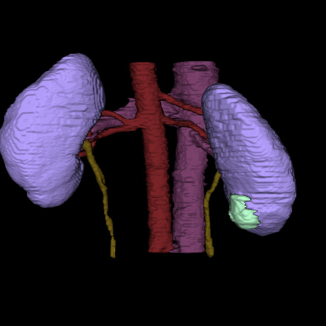
Parse2022
Challenge UserIt is of significant clinical interest to study pulmonary artery structures in the field of medical image analysis. One prerequisite step is to segment pulmonary artery structures from CT with high accuracy and low time-consuming. The segmentation of pulmonary artery structures benefits the quantification of its morphological changes for diagnosis of pulmonary hypertension and thoracic surgery. However, due to the complexity of pulmonary artery topology, automated segmentation of pulmonary artery topology is a challenging task. Besides, the open accessible large-scale CT data with well labeled pulmonary artery are scarce (The large variations of the topological structures from different patients make the annotation an extremely challenging process). The lack of well labeled pulmonary artery hinders the development of automatic pulmonary artery segmentation algorithm. Hence, we try to host the first Pulmonary ARtery SEgmentation challenge in MICCAI 2022 (Named Parse2022) to start a new research topic.

