
Ziyan Huang
ziyanhuang
- China
- Shanghai Jiao Tong University
- Institute of Medical Robotics
- Website
Statistics
- Member for 3 years, 8 months
- 221 challenge submissions
Activity Overview
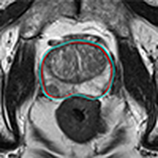
Thyroid Nodule Segmentation and Classification
Challenge UserThe main topic of this TN-SCUI2020 challenge is finding automatic algorithms to accurately classify the thyroid nodules in ultrasound images. It will provide the biggest public dataset of thyroid nodule with over 4500 patient cases from different ages, genders, and were collected using different ultrasound machines. Each ultrasound image is provided with its ground truth class (benign or maglinant) and a detailed delineation of the nodule. This challenge will provide a unique opportunity for participants from different backgrounds (e.g. academia, industry, and government, etc.) to compare their algorithms in an impartial way.
Parse2022
Challenge UserIt is of significant clinical interest to study pulmonary artery structures in the field of medical image analysis. One prerequisite step is to segment pulmonary artery structures from CT with high accuracy and low time-consuming. The segmentation of pulmonary artery structures benefits the quantification of its morphological changes for diagnosis of pulmonary hypertension and thoracic surgery. However, due to the complexity of pulmonary artery topology, automated segmentation of pulmonary artery topology is a challenging task. Besides, the open accessible large-scale CT data with well labeled pulmonary artery are scarce (The large variations of the topological structures from different patients make the annotation an extremely challenging process). The lack of well labeled pulmonary artery hinders the development of automatic pulmonary artery segmentation algorithm. Hence, we try to host the first Pulmonary ARtery SEgmentation challenge in MICCAI 2022 (Named Parse2022) to start a new research topic.
DFUC 2022
Challenge UserDiabetes is a global epidemic affecting around 425 million people and expected to rise to 629 million by 2045. Diabetic Foot Ulcer (DFU) is a severe condition that can result from the disease. The rise of the condition over the last decades is a challenge for healthcare systems. Cases of DFU usually lead to severe conditions that greatly prolongs treatment and result in limb amputation or death. Recent research focuses on creating detection algorithms to monitor their condition to improve patient care and reduce strain on healthcare systems. Work between Manchester Metropolitan University, Lancashire Teaching Hospitals and Manchester University NHS Foundation Trust has created an international repository of up to 11,000 DFU images. Analysis of ulcer regions is a key for DFU management. Delineation of ulcers is time-consuming. With effort from the lead scientists of the UK, US, India and New Zealand, this challenge promotes novel work in DFU segmentation and promote interdisciplinary researcher collaboration.


Multi-site, Multi-Domain Airway Tree Modeling (ATM’22)
Challenge UserAirway segmentation is a crucial step for the analysis of pulmonary diseases including asthma, bronchiectasis, and emphysema. The accurate segmentation based on X-Ray computed tomography (CT) enables the quantitative measurements of airway dimensions and wall thickness, which can reveal the abnormality of patients with chronic obstructive pulmonary disease (COPD). Besides, the extraction of patient-specific airway models from CT images is required for navigatiisted surgery.


ToothFairy: Cone-Beam Computed Tomography Segmentation Challenge
Challenge UserThis is the first edition of the ToothFairy challenge organized by the University of Modena and Reggio Emilia with the collaboration of Raudboud University. This challenge aims at pushing the development of deep learning frameworks to segment the Inferior Alveolar Canal (IAC) by incrementally extending the amount of publicly available 3D-annotated Cone Beam Computed Tomography (CBCT) scans. CBCT modality is becoming increasingly important for treatment planning and diagnosis in implant dentistry and maxillofacial surgery. The three-dimensional information acquired with CBCT can be crucial to plan a vast number of surgical interventions with the aim of preserving noble anatomical structures such as the Inferior Alveolar Canal (IAC), which contains the homonymous nerve (Inferior Alveolar Nerve, IAN). Deep learning models can support medical personnel in surgical planning procedures by providing a voxel-level segmentation of the IAN automatically extracted from CBCT scans.

LNQ2023
Challenge UserAccurate lymph node size estimation is critical for staging cancer patients, initial therapeutic management, and in longitudinal scans, assessing response to therapy. Current standard practice for quantifying lymph node size is based on a variety of criteria that use unidirectional or bidirectional measurements on just one or a few nodes, typically on just one axial slice. But humans have hundreds of lymph nodes, any number of which may be enlarged to various degrees due to disease or immune response. While a normal lymph node may be approximately 5mm in diameter, a diseased lymph node may be several cm in diameter. The mediastinum, the anatomical area between the lungs and around the heart, may contain ten or more lymph nodes, often with three or more enlarged greater than 1cm. Accurate segmentation in 3D would provide more information to evaluate lymph node disease.
SEG.A. - Segmentation of the Aorta
Challenge UserSegmentation, modeling and visualization of the arterial tree are still a challenge in medical image analysis. The main track of this challenge deals with the fully automatic segmentation of the aortic vessel tree in computed tomography images. Optionally, teams can submit tailored solutions for meshing and visualization of the vessel tree.