L Lawliet
L_Lawliet
- United States of America
- Arizona State University
- Computer Science
Statistics
- Member for 2 years, 10 months
- 78 challenge submissions
Activity Overview
IDRiD
Challenge UserThis challenge evaluates automated techniques for analysis of fundus photographs. We target segmentation of retinal lesions like exudates, microaneurysms, and hemorrhages and detection of the optic disc and fovea. Also, we seek grading of fundus images according to the severity level of DR and DME.
Parse2022
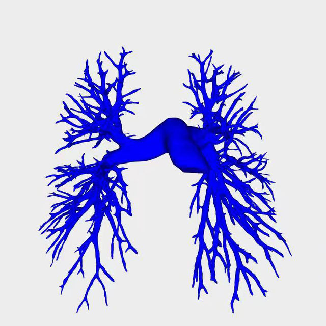
Challenge UserIt is of significant clinical interest to study pulmonary artery structures in the field of medical image analysis. One prerequisite step is to segment pulmonary artery structures from CT with high accuracy and low time-consuming. The segmentation of pulmonary artery structures benefits the quantification of its morphological changes for diagnosis of pulmonary hypertension and thoracic surgery. However, due to the complexity of pulmonary artery topology, automated segmentation of pulmonary artery topology is a challenging task. Besides, the open accessible large-scale CT data with well labeled pulmonary artery are scarce (The large variations of the topological structures from different patients make the annotation an extremely challenging process). The lack of well labeled pulmonary artery hinders the development of automatic pulmonary artery segmentation algorithm. Hence, we try to host the first Pulmonary ARtery SEgmentation challenge in MICCAI 2022 (Named Parse2022) to start a new research topic.
DFUC 2022
Challenge UserDiabetes is a global epidemic affecting around 425 million people and expected to rise to 629 million by 2045. Diabetic Foot Ulcer (DFU) is a severe condition that can result from the disease. The rise of the condition over the last decades is a challenge for healthcare systems. Cases of DFU usually lead to severe conditions that greatly prolongs treatment and result in limb amputation or death. Recent research focuses on creating detection algorithms to monitor their condition to improve patient care and reduce strain on healthcare systems. Work between Manchester Metropolitan University, Lancashire Teaching Hospitals and Manchester University NHS Foundation Trust has created an international repository of up to 11,000 DFU images. Analysis of ulcer regions is a key for DFU management. Delineation of ulcers is time-consuming. With effort from the lead scientists of the UK, US, India and New Zealand, this challenge promotes novel work in DFU segmentation and promote interdisciplinary researcher collaboration.
3D Teeth Scan Segmentation and Labeling Challenge MICCAI2022
Challenge UserComputer-aided design (CAD) tools have become increasingly popular in modern dentistry for highly accurate treatment planning. In particular, in orthodontic CAD systems, advanced intraoral scanners (IOSs) are now widely used as they provide precise digital surface models of the dentition. Such models can dramatically help dentists simulate teeth extraction, move, deletion, and rearrangement and therefore ease the prediction of treatment outcomes. Although IOSs are becoming widespread in clinical dental practice, there are only few contributions on teeth segmentation/labeling available in the literature and no publicly available database. A fundamental issue that appears with IOS data is the ability to reliably segment and identify teeth in scanned observations. Teeth segmentation and labelling is difficult as a result of the inherent similarities between teeth shapes as well as their ambiguous positions on jaws.
Multi-site, Multi-Domain Airway Tree Modeling (ATM’22)
Challenge UserAirway segmentation is a crucial step for the analysis of pulmonary diseases including asthma, bronchiectasis, and emphysema. The accurate segmentation based on X-Ray computed tomography (CT) enables the quantitative measurements of airway dimensions and wall thickness, which can reveal the abnormality of patients with chronic obstructive pulmonary disease (COPD). Besides, the extraction of patient-specific airway models from CT images is required for navigatiisted surgery.

