Your mugshot

Varun Teja Gadde

gaddevarunteja

  •  India
  •  Vignan
  •  Development Department
Statistics
  • Member for 2 years, 3 months

Activity Overview

LOLA11 Logo
LOLA11
Challenge User

The goal of LOLA11 (LObe and Lung Analysis 2011) is to compare methods for (semi-)automatic segmentation of the lungs and lobes from chest computed tomography scans. Any team, whether from academia or industry, can join.

PROMISE12 Logo
PROMISE12
Challenge User

The goal of this challenge is to compare interactive and (semi)-automatic segmentation algorithms for MRI of the prostate.

CAMELYON17 Logo
CAMELYON17
Challenge User

Automated detection and classification of breast cancer metastases in whole-slide images of histological lymph node sections. This task has high clinical relevance and would normally require extensive microscopic assessment by pathologists.

ICIAR2018-Challenge Logo
ICIAR 2018
Challenge User

Can you develop a method for automatic detection of cancerous regions in breast cancer histology images?

SLIVER07 Logo
SLIVER07
Challenge User

The goal of this competition is to compare different algorithms to segment the liver from clinical 3D computed tomography (CT) scans.

drive Logo
DRIVE
Challenge User

Develop a system to automatically segment vessels in human retina fundus images.

REFUGE Logo
REFUGE
Challenge User

The goal of the Retinal Fundus Glaucoma Challenge (REFUGE) is to evaluate and compare automated algorithms for glaucoma detection and optic disc/cup segmentation on a common dataset of retinal fundus images.

PROSTATEx Logo
PROSTATEx
Challenge User

Classification of clinical significance of prostate lesions using multi-parametric MRI data

HC18 Logo
HC18
Challenge User

Automated measurement of fetal head circumference using 2D ultrasound images

ANHIR Logo
ANHIR
Challenge User

The challenge focuses on comparing the accuracy (using manually annotated landmarks) and the approximate speed of automatic non-linear registration methods for aligning microscopy images of multi-stained histology tissue samples.

BreastPathQ Logo
BreastPathQ: Cancer Cellularity Challenge 2019
Challenge User

SPIE-AAPM-NCI BreastPathQ:Cancer Circularity Challenge 2019: Participants will be tasked to develop an automated method for analyzing histology patches extracted from whole slide images and assign a score reflecting cancer cellularity for tumor burden assessment in each.
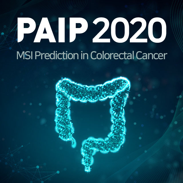
CHAOS Logo
CHAOS
Challenge User

In this challenge, you segment the liver in CT data, and segment liver, spleen, and kidneys in MRI data.

ACDC-LungHP Logo
ACDC-LungHP
Challenge User

Automatic Cancer Detection and Classification in Whole-slide Lung Histopathology

PALM Logo
PALM
Challenge User

The Pathologic Myopia Challenge (PALM) focuses on the investigation and development of algorithms associated with the diagnosis of Pathological Myopia (PM) and segmentation of lesions in fundus photos from PM patients.

Decathlon-10 Logo
Decathlon
Challenge User

The Medical Segmentation Decathlon challenge tests the generalisability of machine learning algorithms when applied to 10 different semantic segmentation task.

LYON19 Logo
LYON19
Challenge User

Automatic Lymphocyte detection in IHC stained specimens.

KiTS19 Logo
KiTS19
Challenge User

2019 Kidney and Kidney Tumor Segmentation Challenge

PAIP2019 Logo
PAIP 2019
Challenge User

PAIP2019: Liver Cancer Segmentation Task 1: Liver Cancer Segmentation Task 2: Viable Tumor Burden Estimation

patchcamelyon Logo
PatchCamelyon
Challenge User

PatchCamelyon is a new and challenging image classification dataset of 327.680 color images (96 x 96px) extracted from histopathology images of the CAMELYON16 challenge. The goal is to detect breast cancer metastasis in lymph nodes.

AGE Logo
AGE
Challenge User

ANGLE CLOSURE GLAUCOMA EVALUATION CHALLENGE

AMD Logo
iChallenge-AMD
Challenge User

Age-related Macular Degeneration Challenge

odir2019 Logo
ODIR-2019
Challenge User

北京大学国际眼底图像智能识别竞赛 Peking University International Competition on Ocular Disease Intelligent Recognition

LYSTO Logo
Lymphocyte Assessment Hackathon
Challenge User

Lymphocyte Assessment Hackathon in conjunction with the MICCAI COMPAY 2019 Workshop on Computational Pathology

LNDb Logo
LNDb Challenge
Challenge User

Lung cancer screening and Fleischner follow-up determination in chest CT through nodule detection, segmentation and characterization

PAIP2020 Logo
PAIP2020
Challenge User

Built on the success of its predecessor, PAIP2020 is the second challenge organized by the Pathology AI Platform (PAIP) and the Seoul National University Hospital (SNUH). PAIP2020 will proceed to not only detect whole tumor areas in colorectal cancers but also to classify their molecular subtypes, which will lead to characterization of their heterogeneity with respect to prognoses and therapeutic responses. All participants should predict one of the molecular carcinogenesis pathways, i.e., microsatellite instability(MSI) in colorectal cancer, by performing digital image analysis without clinical tests. This task has a high clinical relevance as the currently used procedure requires an extensive microscopic assessment by pathologists. Therefore, those automated algorithms would reduce the workload of pathologists as a diagnostic assistance.

RibFrac Logo
RibFrac
Challenge User

Rib Fracture Detection and Classification Challenge: A large-scale benchmark of 660 CT scans with ~5,000 rib fractures (around 80Gb)

Learn2Reg Logo
Learn2Reg
Challenge User

Challenge on medical image registration addressing: learning from small datasets; estimating large deformations; dealing with multi-modal scans; and learning from noisy annotations

DFU2020 Logo
Diabetic Foot Ulcer Challenge 2020
Challenge User

Diabetic Foot Ulcer Challenge 2020

qubiq Logo
QUBIQ
Challenge User

Quantification of Uncertainties in Biomedical Image Segmentation Challenge

lodopab Logo
LoDoPaB-CT
Challenge User

Low-Dose CT reconstruction in the setting of the LoDoPaB-CT dataset.

RIADD Logo
RIADD (ISBI-2021)
Challenge User

Retinal Image Analysis for multi-Disease Detection

MitoEM Logo
MitoEM
Challenge User

Large-scale 3D mitochondria instance segmentation benchmark

covid-segmentation Logo
COVID-19 LUNG CT LESION SEGMENTATION CHALLENGE - 2020
Challenge User

This challenge will create the platform to evaluate emerging methods for the segmentation and quantification of lung lesions caused by SARS-CoV-2 infection from CT images.

SegPC-2021 Logo
SegPC-2021
Challenge User

This challenge is positioned towards robust segmentation of cells which is the first stage to build such a tool for plasma cell cancer, namely, Multiple Myeloma (MM), which is a type of blood cancer.

EndoCV2021 Logo
EndoCV2021
Challenge User

Endoscopy Computer Vision Challenge 2021

crossMoDA Logo
Cross-Modality Domain Adaptation Image Segmentation - 2021
Challenge User

The CrossMoDA challenge 2021 introduces the first large and multi-class medical dataset for unsupervised cross-modality Domain Adaptation.

BrainPTM-2021 Logo
BrainPTM 2021
Challenge User

Brain Pre-surgical Tractography Mapping (BrainPTM) in real clinical scans.

FLARE Logo
FLARE21
Challenge User

Fast and Low GPU memory Abdominal oRgan sEgmentation Challenge

fastPET-LD Logo
fastPET-LD
Challenge User

The purpose of this challenge is the detection of “hot spots” in fast PET scan, that is locations that have an elevated standard uptake value (SUV) and potential clinical significance. Corresponding CT scans are also provided. The ground truth, common to both datasets, was generated by a nuclear medicine expert. It consists of a 3-D segmentation map of the hot spots as well as a text file containing the position and size of 3D cuboid bounding box for each hot spot.

DFU-2021 Logo
DFUC2021
Challenge User

Diabetic Foot Ulcer Challenge 2021

NODE21 Logo
NODE21
Challenge User

NODE21: generate and detect nodules on chest radiographs

WSSS4LUAD Logo
WSSS4LUAD
Challenge User

The WSSS4LUAD dataset contains over 10,000 patches of lung adenocarcinoma from whole slide images from Guangdong Provincial People's Hospital and TCGA with image-level annotations. The goal of this challenge is to perform semantic segmentation for differentiating three important types of tissues in the WSIs of lung adenocarcinoma, including cancerous epithelial region, cancerous stroma region and normal region. Paticipants have to use image-level annotations to give pixel-level prediction.

QUBIQ21 Logo
QUBIQ2021
Challenge User

Quantification of Uncertainties in Biomedical Image Segmentation Challenge 2021

tiger Logo
TIGER
Challenge User

Grand challenge on automate assessment of tumor infiltrating lymphocytes in digital pathology slides of triple negative and Her2-positive breast cancers

STOIC2021 Logo
STOIC2021 - COVID-19 AI Challenge
Challenge User

COVID-19 Artificial Intelligence Challenge: automated diagnosis, and prognostic evaluation based on computed tomography

PI-CAI Logo
The PI-CAI Challenge
Challenge User

Artificial Intelligence and Radiologists at Prostate Cancer Detection in MRI

AIROGS Logo
AIROGS
Challenge User

Artificial Intelligence for RObust Glaucoma Screening Challenge

CoNIC-Challenge Logo
CoNIC 2022
Challenge User

Colon Nuclei Identification and Counting Challenge 2022

3DTeethSeg Logo
3D Teeth Scan Segmentation and Labeling Challenge MICCAI2022
Challenge User

Computer-aided design (CAD) tools have become increasingly popular in modern dentistry for highly accurate treatment planning. In particular, in orthodontic CAD systems, advanced intraoral scanners (IOSs) are now widely used as they provide precise digital surface models of the dentition. Such models can dramatically help dentists simulate teeth extraction, move, deletion, and rearrangement and therefore ease the prediction of treatment outcomes. Although IOSs are becoming widespread in clinical dental practice, there are only few contributions on teeth segmentation/labeling available in the literature and no publicly available database. A fundamental issue that appears with IOS data is the ability to reliably segment and identify teeth in scanned observations. Teeth segmentation and labelling is difficult as a result of the inherent similarities between teeth shapes as well as their ambiguous positions on jaws.