chen junqiang
junqiangchen
- China
- Mediworks
- R&D
- Website
Statistics
- Member for 2 years, 2 months
- 93 challenge submissions
Activity Overview
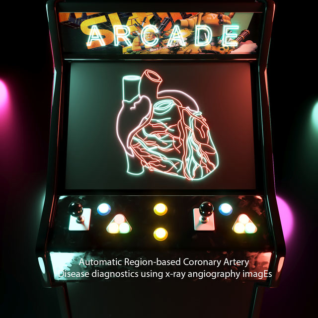
ROCC
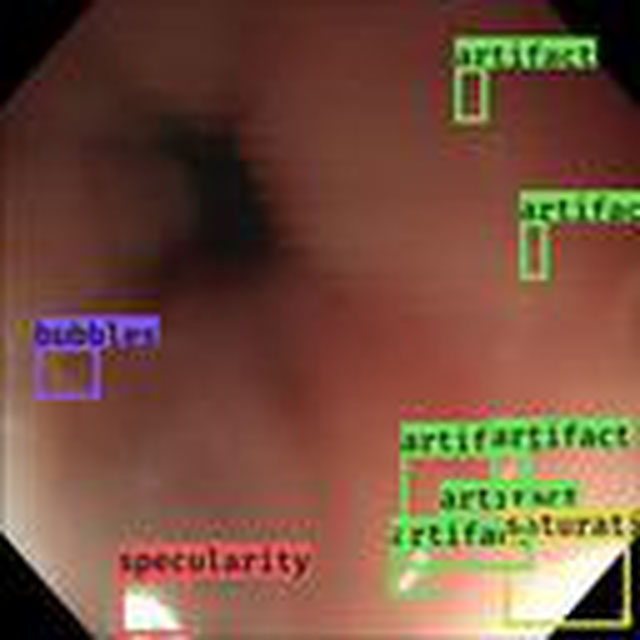
Challenge UserRetinal OCT Classification Challenge (ROCC) is organized as a one day Challenge in conjunction with MVIP2017. The goal of this challenge is to call different automated algorithms that are able to detect DR disease from normal retina on a common dataset of OCT volumes, acquired with Topcon SD-OCT devices.

Gleason2019
Challenge UserMICCAI 2019 Automatic Prostate Gleason Grading Challenge: This challenge aims at the automatic Gleason grading of prostate cancer from H&E-stained histopathology images. This task is of critical importance because Gleason score is a strong prognostic predictor. On the other hand, it is very challenging because of the large degree of heterogeneity in the cellular and glandular patterns associated with each Gleason grade, leading to significant inter-observer variability, even among expert pathologists.


Parse2022
Challenge UserIt is of significant clinical interest to study pulmonary artery structures in the field of medical image analysis. One prerequisite step is to segment pulmonary artery structures from CT with high accuracy and low time-consuming. The segmentation of pulmonary artery structures benefits the quantification of its morphological changes for diagnosis of pulmonary hypertension and thoracic surgery. However, due to the complexity of pulmonary artery topology, automated segmentation of pulmonary artery topology is a challenging task. Besides, the open accessible large-scale CT data with well labeled pulmonary artery are scarce (The large variations of the topological structures from different patients make the annotation an extremely challenging process). The lack of well labeled pulmonary artery hinders the development of automatic pulmonary artery segmentation algorithm. Hence, we try to host the first Pulmonary ARtery SEgmentation challenge in MICCAI 2022 (Named Parse2022) to start a new research topic.


Multi-site, Multi-Domain Airway Tree Modeling (ATM’22)
Challenge UserAirway segmentation is a crucial step for the analysis of pulmonary diseases including asthma, bronchiectasis, and emphysema. The accurate segmentation based on X-Ray computed tomography (CT) enables the quantitative measurements of airway dimensions and wall thickness, which can reveal the abnormality of patients with chronic obstructive pulmonary disease (COPD). Besides, the extraction of patient-specific airway models from CT images is required for navigatiisted surgery.
Shifts Challenge 2022
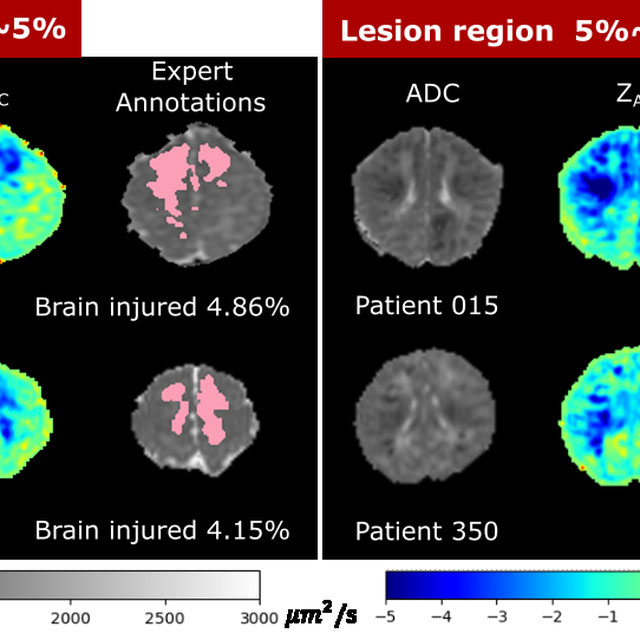
Challenge UserThe goal of the Shifts Challenge 2022 is to raise awareness among the research community about the problems of distributional shift, robustness, and uncertainty estimation, and to identify new solutions to address them. The competition will consist of two new tracks: White Matter Multiple Sclerosis (MS) lesion segmentation in 3D Magnetic Resonance Imaging (MRI) of the brain and Marine cargo vessel power estimation.


ToothFairy: Cone-Beam Computed Tomography Segmentation Challenge
Challenge UserThis is the first edition of the ToothFairy challenge organized by the University of Modena and Reggio Emilia with the collaboration of Raudboud University. This challenge aims at pushing the development of deep learning frameworks to segment the Inferior Alveolar Canal (IAC) by incrementally extending the amount of publicly available 3D-annotated Cone Beam Computed Tomography (CBCT) scans. CBCT modality is becoming increasingly important for treatment planning and diagnosis in implant dentistry and maxillofacial surgery. The three-dimensional information acquired with CBCT can be crucial to plan a vast number of surgical interventions with the aim of preserving noble anatomical structures such as the Inferior Alveolar Canal (IAC), which contains the homonymous nerve (Inferior Alveolar Nerve, IAN). Deep learning models can support medical personnel in surgical planning procedures by providing a voxel-level segmentation of the IAN automatically extracted from CBCT scans.

LNQ2023
Challenge UserAccurate lymph node size estimation is critical for staging cancer patients, initial therapeutic management, and in longitudinal scans, assessing response to therapy. Current standard practice for quantifying lymph node size is based on a variety of criteria that use unidirectional or bidirectional measurements on just one or a few nodes, typically on just one axial slice. But humans have hundreds of lymph nodes, any number of which may be enlarged to various degrees due to disease or immune response. While a normal lymph node may be approximately 5mm in diameter, a diseased lymph node may be several cm in diameter. The mediastinum, the anatomical area between the lungs and around the heart, may contain ten or more lymph nodes, often with three or more enlarged greater than 1cm. Accurate segmentation in 3D would provide more information to evaluate lymph node disease.
SEG.A. - Segmentation of the Aorta
Challenge UserSegmentation, modeling and visualization of the arterial tree are still a challenge in medical image analysis. The main track of this challenge deals with the fully automatic segmentation of the aortic vessel tree in computed tomography images. Optionally, teams can submit tailored solutions for meshing and visualization of the vessel tree.


Light My Cells : Bright Field to Fluorescence Imaging Challenge
Challenge UserJoin the Light My Cells France-Bioimaging challenge! Enhance biology and microscopy by contributing to the development of new image-to-image deep labelling methods. The task: predict the best-focused output images of several fluorescently labelled organelles from label-free transmitted light input images. Dive into the future of imaging with us! 🌐🔬 #LightMyCellsChallenge
Diminished Reality for Emerging Applications in Medicine
Challenge UserThe Diminished Reality for Emerging Applications in Medicine through Inpainting (DREAMING) challenge seeks to pioneer the integration of Diminished Reality (DR) into oral and maxillofacial surgery. While Augmented Reality (AR) has been extensively explored in medicine, DR remains largely uncharted territory. DR involves virtually removing real objects from the environment by replacing them with their background. Recent inpainting methods present an opportunity for real-time DR applications without scene knowledge. DREAMING focuses on implementing such methods to fill obscured regions in surgery scenes with realistic backgrounds, emphasizing the complex facial anatomy and patient diversity. The challenge provides a dataset of synthetic yet photorealistic surgery scenes featuring humans, simulating an operating room setting. Participants are tasked with developing algorithms that seamlessly remove disruptions caused by medical instruments and hands, offering surgeons an unimpeded view of the operative site.

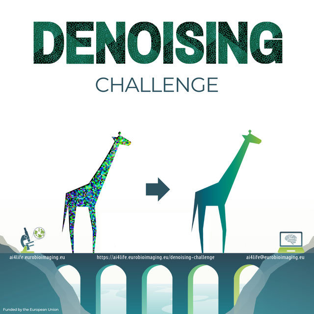
AI4Life Microscopy Denoising Challenge
Challenge UserWellcome to AI4Life-MDC24! In this challenge, we want to focus on an unsupervised denoising of microscopy images. By participating, researchers can contribute to a critical area of scientific research, aiding in interpreting microscopy images and potentially unlocking discoveries in biology and medicine.
ToothFairy2: Multi-Structure Segmentation in CBCT Volumes
Challenge UserThis is the first edition of the ToothFairy challenge organized by the University of Modena and Reggio Emilia with the collaboration of Radboud University Medical Center. The challenge is hosted by grand-challenge and is part of MICCAI2024.