Your mugshot

Ramin Nateghi

ramin

  •  Iran
  •  Shiraz university of Technology SUTECH
  •  Department of Electrical and Electronics Engineering
Statistics
  • Member for 8 years, 10 months
  • 117 challenge submissions
  • 10 algorithms run

Activity Overview

MITOS-ATYPIA-14 Logo
MITOS-ATYPIA-14
Challenge User

MITOS & ATYPIA 14 Contest, hosted by conference ICPR 2014Detection of mitosis and evaluation of nuclear atypia on breast cancer H&E stained images

CAMELYON16 Logo
CAMELYON16
Challenge User

The goal of this challenge is to evaluate new and existing algorithms for automated detection of cancer metastasis in digitized lymph node tissue sections. Two large datasets from both the Radboud University Medical Center and the University Medical Center Utrecht are provided.

LUNA16 Logo
LUNA16
Challenge User

The LUNA16 challenge: automatic nodule detection on chest CT

IDRiD Logo
IDRiD
Challenge User

This challenge evaluates automated techniques for analysis of fundus photographs. We target segmentation of retinal lesions like exudates, microaneurysms, and hemorrhages and detection of the optic disc and fovea. Also, we seek grading of fundus images according to the severity level of DR and DME.
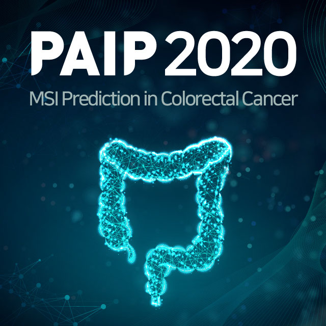
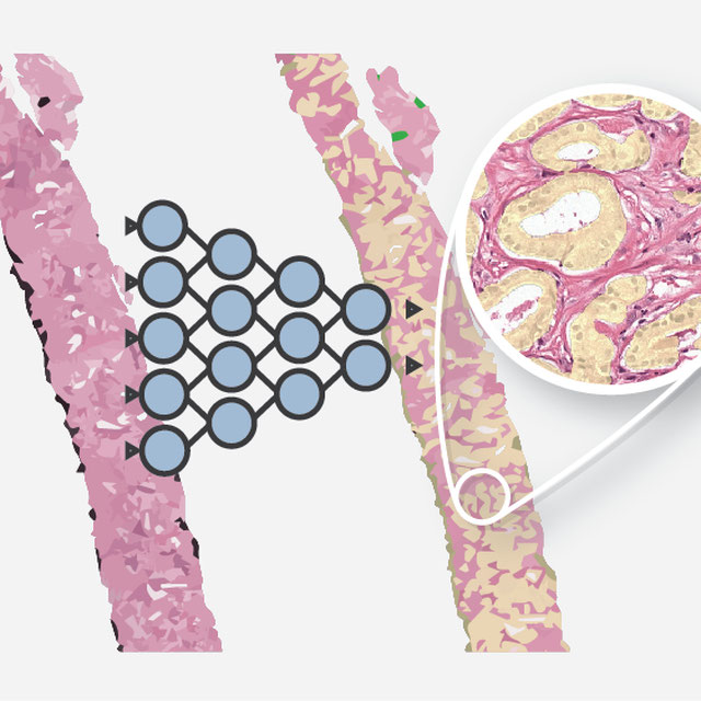
odir2019 Logo
ODIR-2019
Challenge User

北京大学国际眼底图像智能识别竞赛 Peking University International Competition on Ocular Disease Intelligent Recognition

PAIP2020 Logo
PAIP2020
Challenge User

Built on the success of its predecessor, PAIP2020 is the second challenge organized by the Pathology AI Platform (PAIP) and the Seoul National University Hospital (SNUH). PAIP2020 will proceed to not only detect whole tumor areas in colorectal cancers but also to classify their molecular subtypes, which will lead to characterization of their heterogeneity with respect to prognoses and therapeutic responses. All participants should predict one of the molecular carcinogenesis pathways, i.e., microsatellite instability(MSI) in colorectal cancer, by performing digital image analysis without clinical tests. This task has a high clinical relevance as the currently used procedure requires an extensive microscopic assessment by pathologists. Therefore, those automated algorithms would reduce the workload of pathologists as a diagnostic assistance.

RIADD Logo
RIADD (ISBI-2021)
Challenge User

Retinal Image Analysis for multi-Disease Detection

covid-segmentation Logo
COVID-19 LUNG CT LESION SEGMENTATION CHALLENGE - 2020
Challenge User

This challenge will create the platform to evaluate emerging methods for the segmentation and quantification of lung lesions caused by SARS-CoV-2 infection from CT images.

VALDO Logo
Where is VALDO?
Challenge User

Vascular Lesion Detection Challenge at MICCAI 2021

SegPC-2021 Logo
SegPC-2021
Challenge User

This challenge is positioned towards robust segmentation of cells which is the first stage to build such a tool for plasma cell cancer, namely, Multiple Myeloma (MM), which is a type of blood cancer.

Carotid Artery Vessel Wall Segmentation Challenge
Challenge User

To segment the vessel wall of the carotid artery on black-blood MRI images

PAIP2021 Logo
PAIP2021
Challenge User

PAIP 2021 Challenge; Perineural invasion in multiple organ cancer (colon, prostate and pancreatobiliary tract)

NuCLS Logo
NuCLS
Challenge User

Classification, Localization and Segmentation of nuclei in scanned FFPE H&E stained slides of triple-negative breast cancer from The Cancer Genome Atlas. See: Amgad et al. 2021. arXiv:2102.09099 [cs.CV].

BCSegmentation Logo
Breast Cancer Segmentation
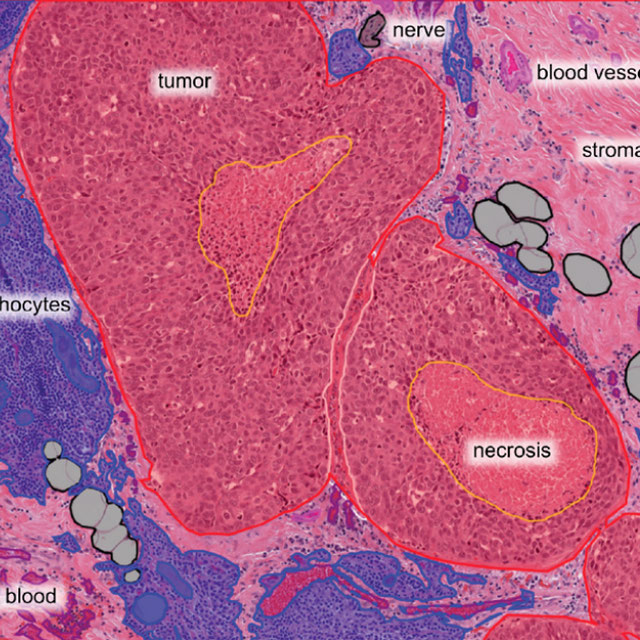
Challenge User

Semantic segmentation of histologic regions in scanned FFPE H&E stained slides of triple-negative breast cancer from The Cancer Genome Atlas. See: Amgad M, Elfandy H, ..., Gutman DA, Cooper LAD. Structured crowdsourcing enables convolutional segmentation of histology images. Bioinformatics. 2019. doi: 10.1093/bioinformatics/btz083
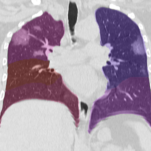
feta Logo
FeTA - Fetal Tissue Annotation Challenge
Challenge User

Fetal Tissue Annotation Challenge

WSSS4LUAD Logo
WSSS4LUAD
Challenge User

The WSSS4LUAD dataset contains over 10,000 patches of lung adenocarcinoma from whole slide images from Guangdong Provincial People's Hospital and TCGA with image-level annotations. The goal of this challenge is to perform semantic segmentation for differentiating three important types of tissues in the WSIs of lung adenocarcinoma, including cancerous epithelial region, cancerous stroma region and normal region. Paticipants have to use image-level annotations to give pixel-level prediction.

QUBIQ21 Logo
QUBIQ2021
Challenge User

Quantification of Uncertainties in Biomedical Image Segmentation Challenge 2021

MIDOG2021 Logo
MIDOG Challenge 2021
Challenge User

Mitosis Domain Generalization Challenge 2021 (part of MICCAI 2021)

tiger Logo
TIGER
Challenge User

Grand challenge on automate assessment of tumor infiltrating lymphocytes in digital pathology slides of triple negative and Her2-positive breast cancers

CXR-COVID19 Logo
Chest XR COVID-19 detection
Challenge User

Build AI models to detect COVID-19 using Chest X-ray images

CoNIC-Challenge Logo
CoNIC 2022
Challenge User

Colon Nuclei Identification and Counting Challenge 2022

BCNB Logo
BCNB
Challenge User

Early Breast Cancer Core-Needle Biopsy WSI Dataset

AGGC22 Logo
AGGC22
Challenge User

ACROBAT Logo
ACROBAT 2023
Challenge User

The ACROBAT challenge aims to advance the development of WSI registration algorithms that can align WSIs of IHC-stained breast cancer tissue sections to corresponding tissue regions that were stained with H&E. All WSIs originate from routine diagnostic workflows.

MIDOG2022 Logo
MItosis DOmain Generalization Challenge 2022
Challenge User

2023PAIP Logo
PAIP 2023: TC prediction in pancreatic and colon cancer
Challenge User

Tumor cellularity prediction in pancreatic cancer (supervised learning) and colon cancer (transfer learning)

OCELOT2023 Logo
OCELOT 2023: Cell Detection from Cell-Tissue Interaction
Challenge User

Gleason Grading of Prostate Biopsies Logo
Gleason Grading of Prostate Biopsies
Algorithm User

Automated Gleason grading of prostate biopsies following the Gleason Grade Group system.

Pulmonary Lobe Segmentation Logo
Pulmonary Lobe Segmentation
Algorithm User

Automatic segmentation of pulmonary lobes on CT scans for patients with COPD or COVID-19.

CORADS-AI Logo
CORADS-AI
Algorithm User

Segments pulmonary lobes and lesions and computes the CORADS and CT Severity Score from a non-contrast CT scan.

Neural Image Compression Logo
Neural Image Compression
Algorithm User

Compresses whole slide images into much smaller volumes

MIDOG FR1 Algorithm Logo
MIDOG FR1 Algorithm
Algorithm Editor