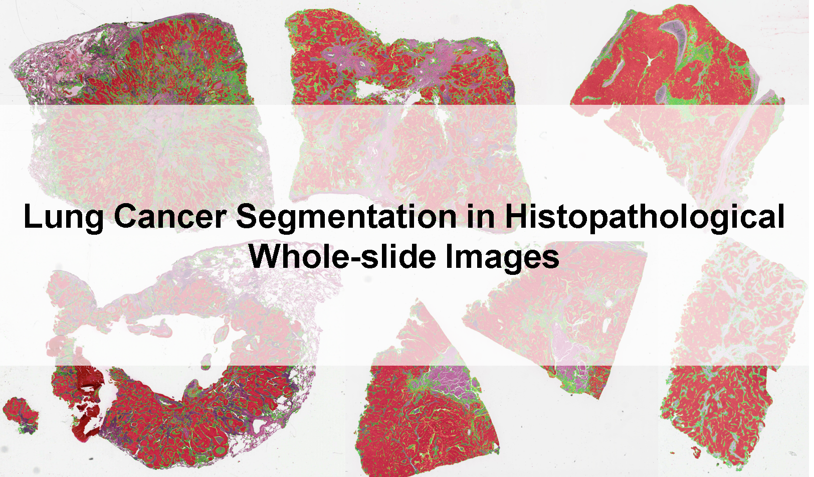
Lung Cancer Segmentation

About
Contact email:
Image Version:
50577ada-46fe-431d-8064-ad11e68c3c2d — May 3, 2022
Summary
The model was trained on ACDC-HP challenge and got 3rd place in post-challenge leaderboard. It was further fine-tuned with data from the TCGA-LUAD and TCGA-LUSC projects.
The input images should be whole-slide images, with formats including 'svs', 'tif' etc. The images should have three channels (RGB) and a pixel spacing of about 0.5μm/pixel.
Mechanism
The model is based on U-Net, and trained with data from ACDC-HP challenge, as well as in-house annotations of TCGA-LUAD and TCGA-LUSC
Interfaces
This algorithm implements all of the following input-output combinations:
Validation and Performance
Left empty by the Algorithm Editors
Uses and Directions
This algorithm was developed for research purposes only.
Warnings
Left empty by the Algorithm Editors
Common Error Messages
Left empty by the Algorithm Editors
Information on this algorithm has been provided by the Algorithm Editors,
following the Model Facts labels guidelines from
Sendak, M.P., Gao, M., Brajer, N. et al.
Presenting machine learning model information to clinical end users with model facts labels.
npj Digit. Med. 3, 41 (2020). 10.1038/s41746-020-0253-3