Spleen Segmentation

About
Editors:


Contact email:
Image Version:
e9694929-e0fd-42af-8fba-318282d55f02 — April 4, 2019
Summary
General information
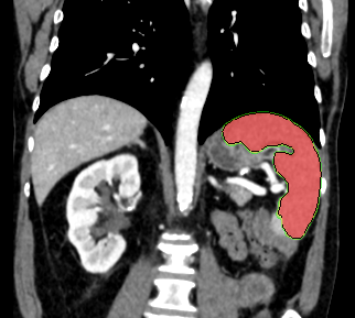
This algorithm fully automatically performs a volumetric segmentation of the spleen on contrast-enhanced thorax-abdomen CT scans. The underlying algorithm is a 3D U-Net model trained and validated on a large set of contrast-enhanced CT thorax abdomen scans from one academic center in the Netherlands. A large proportion of the training scans came from oncological follow-up and hence contained a wide variety of abnormalities and pathology. The algorithm and its validation is described in this publication in Radiology: Artificial Intelligence .
Contact information
For questions about this algorithm, please contact Colin Jacobs.
Mechanism
Left empty by the Algorithm Editors
Interfaces
This algorithm implements all of the following input-output combinations:
Validation and Performance
Left empty by the Algorithm Editors
Uses and Directions
This algorithm was developed for research purposes only.
Warnings
Left empty by the Algorithm Editors
Common Error Messages
Left empty by the Algorithm Editors
Information on this algorithm has been provided by the Algorithm Editors,
following the Model Facts labels guidelines from
Sendak, M.P., Gao, M., Brajer, N. et al.
Presenting machine learning model information to clinical end users with model facts labels.
npj Digit. Med. 3, 41 (2020). 10.1038/s41746-020-0253-3