Your mugshot

Fabian Isensee

Isensee

  •  Germany
  •  German Cancer Research Center (DKFZ)
  •  Medical Image Computing
Statistics
  • Member for 6 years, 3 months
  • 28 challenge submissions

Activity Overview

VESSEL12 Logo
VESSEL12
Challenge User

The VESSEL12 challenge compares methods for automatic (and semi-automatic) segmentation of blood vessels in the lungs from CT images.

PROMISE12 Logo
PROMISE12
Challenge User

The goal of this challenge is to compare interactive and (semi)-automatic segmentation algorithms for MRI of the prostate.

LUNA16 Logo
LUNA16
Challenge User

The LUNA16 challenge: automatic nodule detection on chest CT

CAMELYON17 Logo
CAMELYON17
Challenge User

Automated detection and classification of breast cancer metastases in whole-slide images of histological lymph node sections. This task has high clinical relevance and would normally require extensive microscopic assessment by pathologists.

drive Logo
DRIVE
Challenge User

Develop a system to automatically segment vessels in human retina fundus images.

HC18 Logo
HC18
Challenge User

Automated measurement of fetal head circumference using 2D ultrasound images

CHAOS Logo
CHAOS
Challenge User

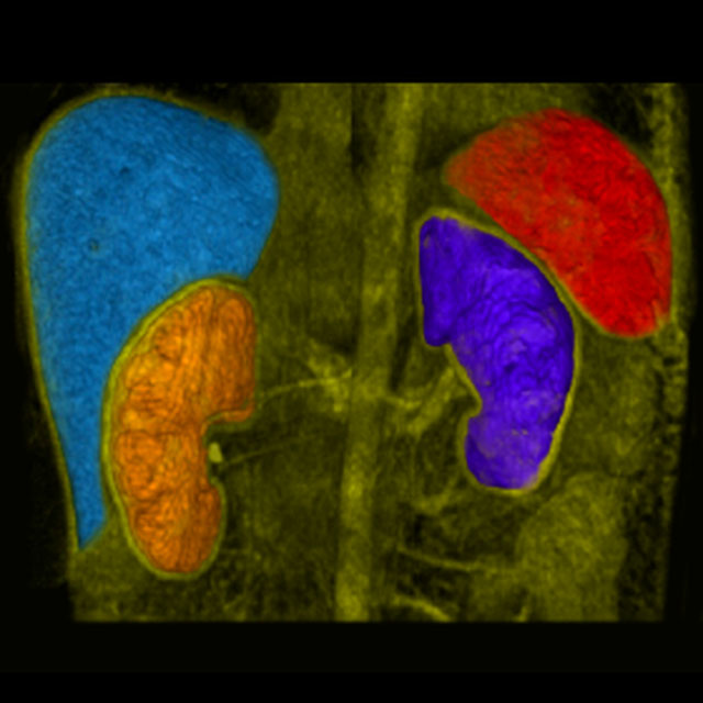
In this challenge, you segment the liver in CT data, and segment liver, spleen, and kidneys in MRI data.

PALM Logo
PALM
Challenge User

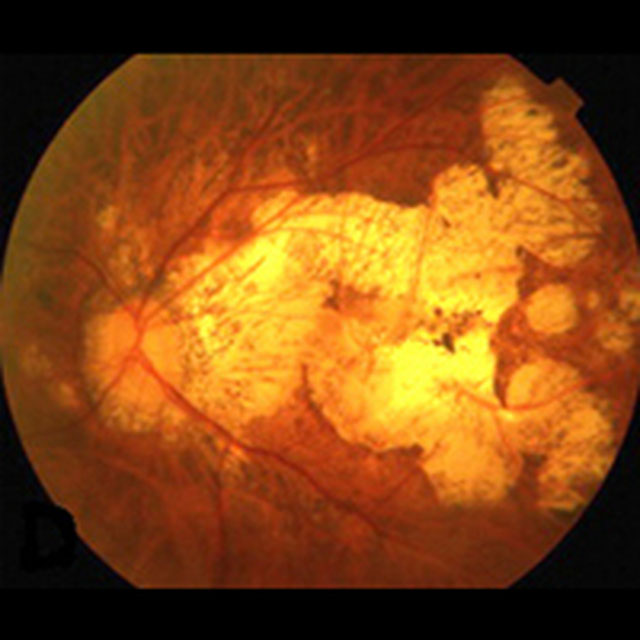
The Pathologic Myopia Challenge (PALM) focuses on the investigation and development of algorithms associated with the diagnosis of Pathological Myopia (PM) and segmentation of lesions in fundus photos from PM patients.

Decathlon-10 Logo
Decathlon
Challenge User

The Medical Segmentation Decathlon challenge tests the generalisability of machine learning algorithms when applied to 10 different semantic segmentation task.

KiTS19 Logo
KiTS19
Challenge User

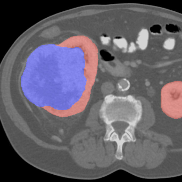
2019 Kidney and Kidney Tumor Segmentation Challenge

VerSe2019 Logo
VerSe`19
Challenge User

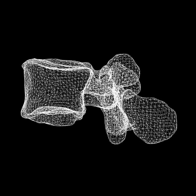
Vertebrae labelling and segmentation on a spine dataset on an unprecedented 150 CT scans with voxel-level vertebral annotations.

StructSeg2019 Logo
StructSeg2019
Challenge User

Welcome to Automatic Structure Segmentation for Radiotherapy Planning Challenge 2019. This competition is part of the MICCAI 2019 Challenge.

EndoCV Logo
EndoCV2020
Challenge User

Endoscopy computer vision challenge (EndoCV2020) introduces two core sub-themes in endoscopy: 1) artefact detection and segmentation (EAD2020) and 2) disease detection and segmentation (EDD2020).

VerSe2020 Logo
VerSe'20
Challenge User

Vertebrae labelling and segmentation on a multi-centre, multi-scanner, and anatomically-diverse CT dataset.

covid-segmentation Logo
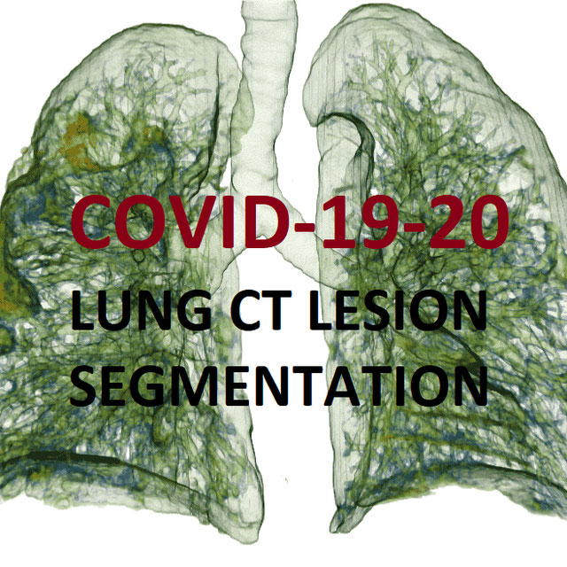
COVID-19 LUNG CT LESION SEGMENTATION CHALLENGE - 2020
Challenge User

This challenge will create the platform to evaluate emerging methods for the segmentation and quantification of lung lesions caused by SARS-CoV-2 infection from CT images.

SegPC-2021 Logo
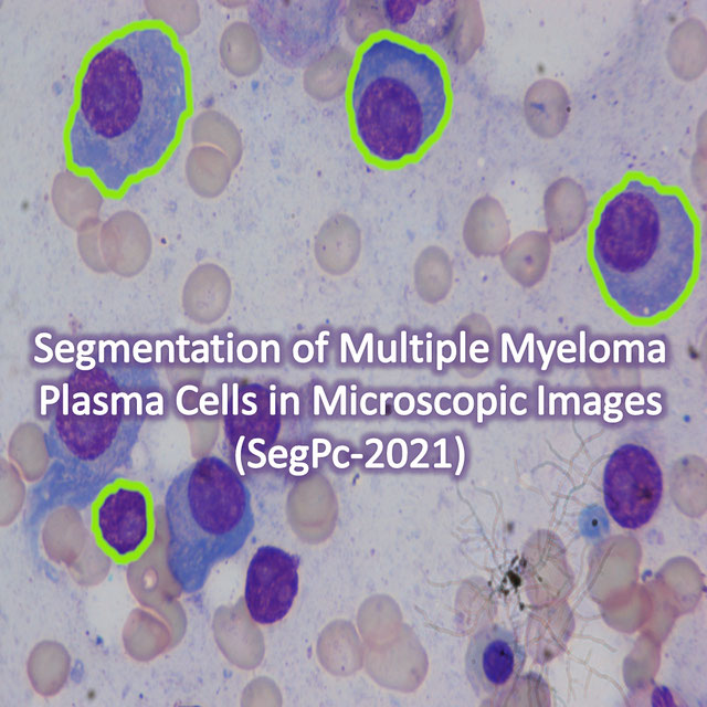
SegPC-2021
Challenge User

This challenge is positioned towards robust segmentation of cells which is the first stage to build such a tool for plasma cell cancer, namely, Multiple Myeloma (MM), which is a type of blood cancer.

PAIP2021 Logo
PAIP2021
Challenge User

PAIP 2021 Challenge; Perineural invasion in multiple organ cancer (colon, prostate and pancreatobiliary tract)

FLARE Logo
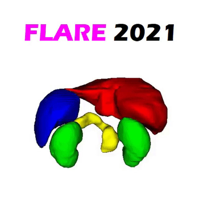
FLARE21
Challenge User

Fast and Low GPU memory Abdominal oRgan sEgmentation Challenge

kipa22 Logo
KiPA22 (Regular Challenge)
Challenge User

The challenge is aimed to segment kidney, renal tumors, arteries, and veins from computed tomography angiography (CTA) images in one inference.

Parse2022 Logo
Parse2022
Challenge User

It is of significant clinical interest to study pulmonary artery structures in the field of medical image analysis. One prerequisite step is to segment pulmonary artery structures from CT with high accuracy and low time-consuming. The segmentation of pulmonary artery structures benefits the quantification of its morphological changes for diagnosis of pulmonary hypertension and thoracic surgery. However, due to the complexity of pulmonary artery topology, automated segmentation of pulmonary artery topology is a challenging task. Besides, the open accessible large-scale CT data with well labeled pulmonary artery are scarce (The large variations of the topological structures from different patients make the annotation an extremely challenging process). The lack of well labeled pulmonary artery hinders the development of automatic pulmonary artery segmentation algorithm. Hence, we try to host the first Pulmonary ARtery SEgmentation challenge in MICCAI 2022 (Named Parse2022) to start a new research topic.

3DTeethSeg Logo
3D Teeth Scan Segmentation and Labeling Challenge MICCAI2022
Challenge User

Computer-aided design (CAD) tools have become increasingly popular in modern dentistry for highly accurate treatment planning. In particular, in orthodontic CAD systems, advanced intraoral scanners (IOSs) are now widely used as they provide precise digital surface models of the dentition. Such models can dramatically help dentists simulate teeth extraction, move, deletion, and rearrangement and therefore ease the prediction of treatment outcomes. Although IOSs are becoming widespread in clinical dental practice, there are only few contributions on teeth segmentation/labeling available in the literature and no publicly available database. A fundamental issue that appears with IOS data is the ability to reliably segment and identify teeth in scanned observations. Teeth segmentation and labelling is difficult as a result of the inherent similarities between teeth shapes as well as their ambiguous positions on jaws.

autoPET Logo
autoPET
Challenge User

Automatic lesion segmentation in whole-body FDG-PET/CT

AMOS22 Logo
Multi-Modality Abdominal Multi-Organ Segmentation Challenge 2022
Challenge User

autoPET-II Logo
autoPET-II
Challenge User

Automated Lesion Segmentation in PET/CT - Domain Generalization

MultiCenterAorta Logo
SEG.A. - Segmentation of the Aorta
Challenge User

Segmentation, modeling and visualization of the arterial tree are still a challenge in medical image analysis. The main track of this challenge deals with the fully automatic segmentation of the aortic vessel tree in computed tomography images. Optionally, teams can submit tailored solutions for meshing and visualization of the vessel tree.

TopCoW23 Logo
Topology-Aware Anatomical Segmentation of the Circle of Willis
Challenge User

Segment the Circle of Willis (CoW) vessel components for both CTA and MRA

SegRap2023 Logo
SegRap 2023
Challenge User

A segmentation challenge with 200 patients (two modalities of CT images, 45 OARs and 2 GTVs).

Balaitous Logo
Balaitous
Algorithm User

A deep learning model to estimate COVID disease and severity from a CT scan