hao li
lihaossu
- South Korea
- Hutom
- MIAI
- Website
Statistics
- Member for 5 years, 11 months
- 1 challenge submissions
Activity Overview


Multi-site, Multi-Domain Airway Tree Modeling (ATM’22)
Challenge UserAirway segmentation is a crucial step for the analysis of pulmonary diseases including asthma, bronchiectasis, and emphysema. The accurate segmentation based on X-Ray computed tomography (CT) enables the quantitative measurements of airway dimensions and wall thickness, which can reveal the abnormality of patients with chronic obstructive pulmonary disease (COPD). Besides, the extraction of patient-specific airway models from CT images is required for navigatiisted surgery.

LNQ2023
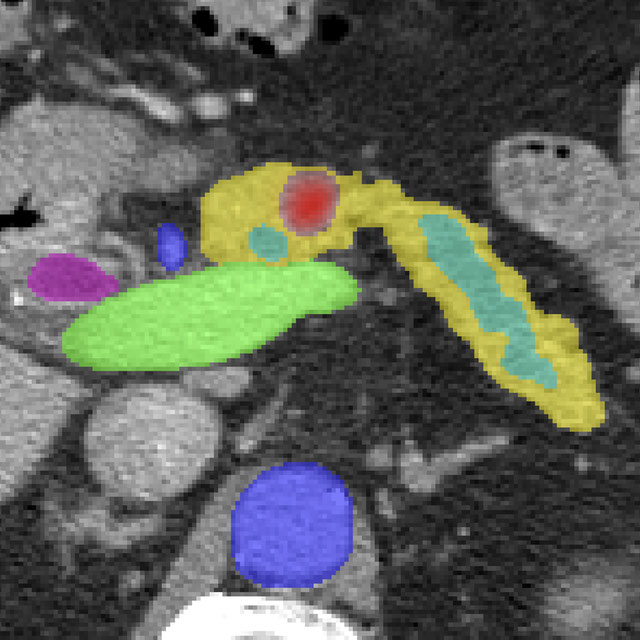
Challenge UserAccurate lymph node size estimation is critical for staging cancer patients, initial therapeutic management, and in longitudinal scans, assessing response to therapy. Current standard practice for quantifying lymph node size is based on a variety of criteria that use unidirectional or bidirectional measurements on just one or a few nodes, typically on just one axial slice. But humans have hundreds of lymph nodes, any number of which may be enlarged to various degrees due to disease or immune response. While a normal lymph node may be approximately 5mm in diameter, a diseased lymph node may be several cm in diameter. The mediastinum, the anatomical area between the lungs and around the heart, may contain ten or more lymph nodes, often with three or more enlarged greater than 1cm. Accurate segmentation in 3D would provide more information to evaluate lymph node disease.