Vivek Kumar Singh
vivek23
- Spain
- Universitat Rovira I Virgili
- Department of Engineering Informatics and Mathematics
Statistics
- Member for 5 years, 10 months
- 117 challenge submissions
Activity Overview
ROCC
Challenge UserRetinal OCT Classification Challenge (ROCC) is organized as a one day Challenge in conjunction with MVIP2017. The goal of this challenge is to call different automated algorithms that are able to detect DR disease from normal retina on a common dataset of OCT volumes, acquired with Topcon SD-OCT devices.
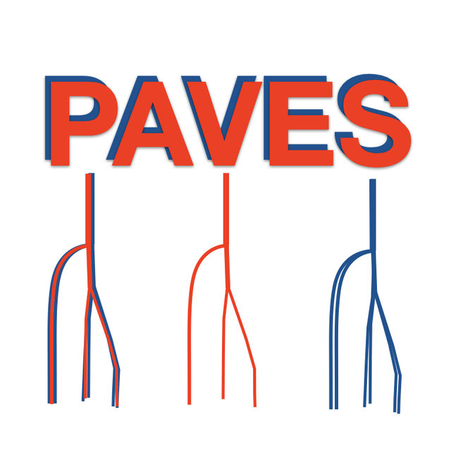
PAVES
Challenge UserPeripheral Artery:Vein Enhanced Segmentation (PAVES) is the challenge focussed on providing easily interpretable and relevant images that can be readily understood by clinicians (vascular interventional radiologists & vascular surgeons) from MRA datasets where the venous and arterial vasculature may be equally enhanced. The setting is lower limb arterial occlusive disease where imaging of the below knee arterial vasculature is critical in planning limb salvage interventions. However, the competing demands of the high spatial resolution needed to image small vessels versus imaging time constraints where there is often a very short arteriovenous transit time for contrast passage form arterial to venous compartments makes imaging challenging. While dynamic MRA techniques can usually allow arterial imaging without venous ‘contamination’ these necessarily sacrifice spatial resolution.

Thyroid Nodule Segmentation and Classification
Challenge UserThe main topic of this TN-SCUI2020 challenge is finding automatic algorithms to accurately classify the thyroid nodules in ultrasound images. It will provide the biggest public dataset of thyroid nodule with over 4500 patient cases from different ages, genders, and were collected using different ultrasound machines. Each ultrasound image is provided with its ground truth class (benign or maglinant) and a detailed delineation of the nodule. This challenge will provide a unique opportunity for participants from different backgrounds (e.g. academia, industry, and government, etc.) to compare their algorithms in an impartial way.
SARAS-ESAD
Challenge UserThis challenge is part of Medical Imaging with Deep Learning conference, 2020. The conference is held between 6 ‑ 8 July 2020 in Montréal. The SARAS (Smart Autonomous Robotic Assistant Surgeon) EU consortium, www.saras-project.eu, is working towards replacing the assistant surgeon in MIS with two assistive robotic arms. To accomplish that, an artificial intelligence based system is required which not only can understand the complete surgical scene but also detect the actions being performed by the main surgeon. This information can later be used infer the response required from the autonomous assistant surgeon.
CT diagnosis of COVID-19
Challenge UserCoronavirus disease 2019 (COVID-19) has infected more than 1.3 million individuals all over the world and caused more than 106,000 deaths. One major hurdle in controlling the spreading of this disease is the inefficiency and shortage of medical tests. To mitigate the inefficiency and shortage of existing tests for COVID-19, we propose this competition to encourage the development of effective Deep Learning techniques to diagnose COVID-19 based on CT images. The problem we want to solve is to classify each CT image into positive COVID-19 (the image has clinical findings of COVID-19) or negative COVID-19 ( the image does not have clinical findings of COVID-19). It’s a binary classification problem based on CT images.
A-AFMA
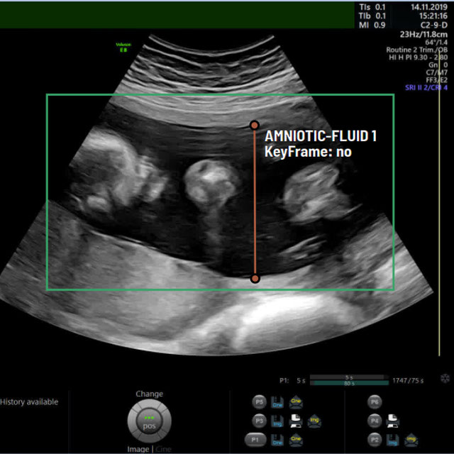
Challenge UserPrenatal ultrasound (US) measurement of amniotic fluid is an important part of fetal surveillance as it provides a non-invasive way of assessing whether there is oligohydramnios (insufficient amniotic fluid) and polyhydramnios (excess amniotic fluid), which are associated with numerous problems both during pregnancy and after birth. In this Image Analysis Challenge, we aim to attract attention from the image analysis community to work on the problem of automated measurement of the MVP using the predefined ultrasound video clip based on a linear-sweep protocol [1]. We define two tasks. The first task is to automatically detect amniotic fluid and the maternal bladder. The second task is to identify the appropriate points for MVP measurement given the selected frame of the video clip, and calculate the length of the connected line between these points. The data was collected from women in the second trimester of pregnancy, as part of the PURE study at the John Radcliffe Hospital in Oxford, UK.

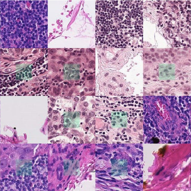
Breast Cancer Segmentation
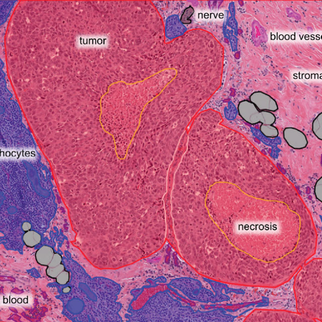
Challenge UserSemantic segmentation of histologic regions in scanned FFPE H&E stained slides of triple-negative breast cancer from The Cancer Genome Atlas. See: Amgad M, Elfandy H, ..., Gutman DA, Cooper LAD. Structured crowdsourcing enables convolutional segmentation of histology images. Bioinformatics. 2019. doi: 10.1093/bioinformatics/btz083
fastPET-LD
Challenge UserThe purpose of this challenge is the detection of “hot spots” in fast PET scan, that is locations that have an elevated standard uptake value (SUV) and potential clinical significance. Corresponding CT scans are also provided. The ground truth, common to both datasets, was generated by a nuclear medicine expert. It consists of a 3-D segmentation map of the hot spots as well as a text file containing the position and size of 3D cuboid bounding box for each hot spot.

WSSS4LUAD
Challenge UserThe WSSS4LUAD dataset contains over 10,000 patches of lung adenocarcinoma from whole slide images from Guangdong Provincial People's Hospital and TCGA with image-level annotations. The goal of this challenge is to perform semantic segmentation for differentiating three important types of tissues in the WSIs of lung adenocarcinoma, including cancerous epithelial region, cancerous stroma region and normal region. Paticipants have to use image-level annotations to give pixel-level prediction.
Parse2022
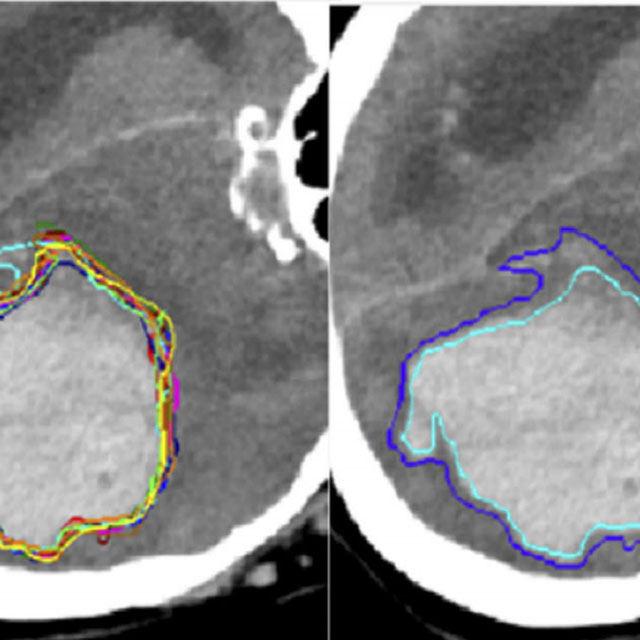
Challenge UserIt is of significant clinical interest to study pulmonary artery structures in the field of medical image analysis. One prerequisite step is to segment pulmonary artery structures from CT with high accuracy and low time-consuming. The segmentation of pulmonary artery structures benefits the quantification of its morphological changes for diagnosis of pulmonary hypertension and thoracic surgery. However, due to the complexity of pulmonary artery topology, automated segmentation of pulmonary artery topology is a challenging task. Besides, the open accessible large-scale CT data with well labeled pulmonary artery are scarce (The large variations of the topological structures from different patients make the annotation an extremely challenging process). The lack of well labeled pulmonary artery hinders the development of automatic pulmonary artery segmentation algorithm. Hence, we try to host the first Pulmonary ARtery SEgmentation challenge in MICCAI 2022 (Named Parse2022) to start a new research topic.
DFUC 2022
Challenge UserDiabetes is a global epidemic affecting around 425 million people and expected to rise to 629 million by 2045. Diabetic Foot Ulcer (DFU) is a severe condition that can result from the disease. The rise of the condition over the last decades is a challenge for healthcare systems. Cases of DFU usually lead to severe conditions that greatly prolongs treatment and result in limb amputation or death. Recent research focuses on creating detection algorithms to monitor their condition to improve patient care and reduce strain on healthcare systems. Work between Manchester Metropolitan University, Lancashire Teaching Hospitals and Manchester University NHS Foundation Trust has created an international repository of up to 11,000 DFU images. Analysis of ulcer regions is a key for DFU management. Delineation of ulcers is time-consuming. With effort from the lead scientists of the UK, US, India and New Zealand, this challenge promotes novel work in DFU segmentation and promote interdisciplinary researcher collaboration.