
pengxin yu
smilenaxx
- China
- Infervision
- AI-Lab
Statistics
- Member for 5 years, 10 months
- 86 challenge submissions
- 15 algorithms run
Activity Overview


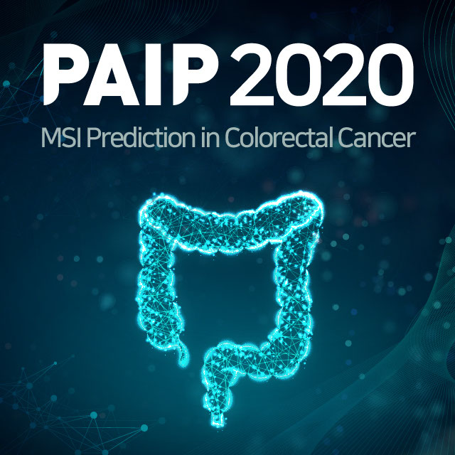
PAIP2020
Challenge UserBuilt on the success of its predecessor, PAIP2020 is the second challenge organized by the Pathology AI Platform (PAIP) and the Seoul National University Hospital (SNUH). PAIP2020 will proceed to not only detect whole tumor areas in colorectal cancers but also to classify their molecular subtypes, which will lead to characterization of their heterogeneity with respect to prognoses and therapeutic responses. All participants should predict one of the molecular carcinogenesis pathways, i.e., microsatellite instability(MSI) in colorectal cancer, by performing digital image analysis without clinical tests. This task has a high clinical relevance as the currently used procedure requires an extensive microscopic assessment by pathologists. Therefore, those automated algorithms would reduce the workload of pathologists as a diagnostic assistance.
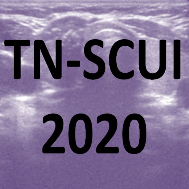
Thyroid Nodule Segmentation and Classification
Challenge UserThe main topic of this TN-SCUI2020 challenge is finding automatic algorithms to accurately classify the thyroid nodules in ultrasound images. It will provide the biggest public dataset of thyroid nodule with over 4500 patient cases from different ages, genders, and were collected using different ultrasound machines. Each ultrasound image is provided with its ground truth class (benign or maglinant) and a detailed delineation of the nodule. This challenge will provide a unique opportunity for participants from different backgrounds (e.g. academia, industry, and government, etc.) to compare their algorithms in an impartial way.


Parse2022
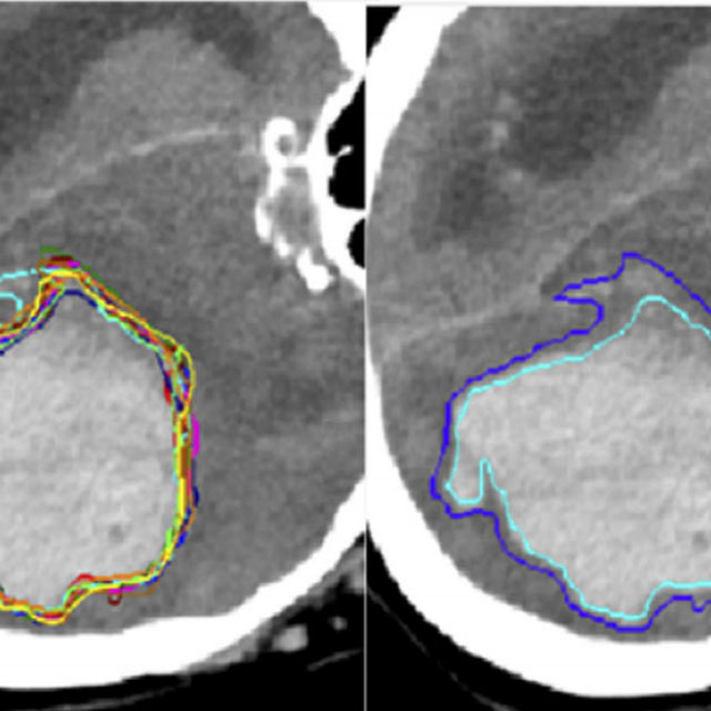
Challenge UserIt is of significant clinical interest to study pulmonary artery structures in the field of medical image analysis. One prerequisite step is to segment pulmonary artery structures from CT with high accuracy and low time-consuming. The segmentation of pulmonary artery structures benefits the quantification of its morphological changes for diagnosis of pulmonary hypertension and thoracic surgery. However, due to the complexity of pulmonary artery topology, automated segmentation of pulmonary artery topology is a challenging task. Besides, the open accessible large-scale CT data with well labeled pulmonary artery are scarce (The large variations of the topological structures from different patients make the annotation an extremely challenging process). The lack of well labeled pulmonary artery hinders the development of automatic pulmonary artery segmentation algorithm. Hence, we try to host the first Pulmonary ARtery SEgmentation challenge in MICCAI 2022 (Named Parse2022) to start a new research topic.
Multi-site, Multi-Domain Airway Tree Modeling (ATM’22)
Challenge UserAirway segmentation is a crucial step for the analysis of pulmonary diseases including asthma, bronchiectasis, and emphysema. The accurate segmentation based on X-Ray computed tomography (CT) enables the quantitative measurements of airway dimensions and wall thickness, which can reveal the abnormality of patients with chronic obstructive pulmonary disease (COPD). Besides, the extraction of patient-specific airway models from CT images is required for navigatiisted surgery.