Your mugshot

tan zhengbo

tanbobo

  •  China
  •  SiChuan University
  •  College of Materials and Engineering
Statistics
  • Member for 4 years, 7 months

Activity Overview

VESSEL12 Logo
VESSEL12
Challenge User

The VESSEL12 challenge compares methods for automatic (and semi-automatic) segmentation of blood vessels in the lungs from CT images.

CRASS Logo
CRASS
Challenge User

CRASS stands for Chest Radiograph Anatomical Structure Segmentation. The challenge currently invites participants to send in results for clavicle segmentation algorithms.

CAUSE07 Logo
CAUSE07
Challenge User

The goal of CAUSE07 is to compare different algorithms to segment the caudate nucleaus from brain MRI scans.

ICIAR2018-Challenge Logo
ICIAR 2018
Challenge User

Can you develop a method for automatic detection of cancerous regions in breast cancer histology images?

ROCC Logo
ROCC
Challenge User

Retinal OCT Classification Challenge (ROCC) is organized as a one day Challenge in conjunction with MVIP2017. The goal of this challenge is to call different automated algorithms that are able to detect DR disease from normal retina on a common dataset of OCT volumes, acquired with Topcon SD-OCT devices.

REFUGE Logo
REFUGE
Challenge User

The goal of the Retinal Fundus Glaucoma Challenge (REFUGE) is to evaluate and compare automated algorithms for glaucoma detection and optic disc/cup segmentation on a common dataset of retinal fundus images.

HC18 Logo
HC18
Challenge User

Automated measurement of fetal head circumference using 2D ultrasound images

PALM Logo
PALM
Challenge User

The Pathologic Myopia Challenge (PALM) focuses on the investigation and development of algorithms associated with the diagnosis of Pathological Myopia (PM) and segmentation of lesions in fundus photos from PM patients.

VerSe2019 Logo
VerSe`19
Challenge User

Vertebrae labelling and segmentation on a spine dataset on an unprecedented 150 CT scans with voxel-level vertebral annotations.

DigestPath2019 Logo
DigestPath2019
Challenge User

Welcome to Digestive-System Pathological Detection and Segmentation Challenge 2019. This competition is part of the MICCAI 2019 Challenge.

odir2019 Logo
ODIR-2019
Challenge User

北京大学国际眼底图像智能识别竞赛 Peking University International Competition on Ocular Disease Intelligent Recognition

ECDP2020 Logo
HEROHE
Challenge User

Unlike previous challenges, this proposes to find an image analysis algorithm to identify HER2-positive from HER2-negative breast cancer specimens evaluating only the morphological features present on the HE slide, without the staining patterns of IHC.

LNDb Logo
LNDb Challenge
Challenge User

Lung cancer screening and Fleischner follow-up determination in chest CT through nodule detection, segmentation and characterization

PAIP2020 Logo
PAIP2020
Challenge User

Built on the success of its predecessor, PAIP2020 is the second challenge organized by the Pathology AI Platform (PAIP) and the Seoul National University Hospital (SNUH). PAIP2020 will proceed to not only detect whole tumor areas in colorectal cancers but also to classify their molecular subtypes, which will lead to characterization of their heterogeneity with respect to prognoses and therapeutic responses. All participants should predict one of the molecular carcinogenesis pathways, i.e., microsatellite instability(MSI) in colorectal cancer, by performing digital image analysis without clinical tests. This task has a high clinical relevance as the currently used procedure requires an extensive microscopic assessment by pathologists. Therefore, those automated algorithms would reduce the workload of pathologists as a diagnostic assistance.

RibFrac Logo
RibFrac
Challenge User

Rib Fracture Detection and Classification Challenge: A large-scale benchmark of 660 CT scans with ~5,000 rib fractures (around 80Gb)

TN-SCUI2020 Logo
Thyroid Nodule Segmentation and Classification
Challenge User

The main topic of this TN-SCUI2020 challenge is finding automatic algorithms to accurately classify the thyroid nodules in ultrasound images. It will provide the biggest public dataset of thyroid nodule with over 4500 patient cases from different ages, genders, and were collected using different ultrasound machines. Each ultrasound image is provided with its ground truth class (benign or maglinant) and a detailed delineation of the nodule. This challenge will provide a unique opportunity for participants from different backgrounds (e.g. academia, industry, and government, etc.) to compare their algorithms in an impartial way.
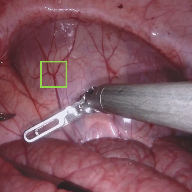
SARAS-ESAD
Challenge User

This challenge is part of Medical Imaging with Deep Learning conference, 2020. The conference is held between 6 ‑ 8 July 2020 in Montréal. The SARAS (Smart Autonomous Robotic Assistant Surgeon) EU consortium, www.saras-project.eu, is working towards replacing the assistant surgeon in MIS with two assistive robotic arms. To accomplish that, an artificial intelligence based system is required which not only can understand the complete surgical scene but also detect the actions being performed by the main surgeon. This information can later be used infer the response required from the autonomous assistant surgeon.
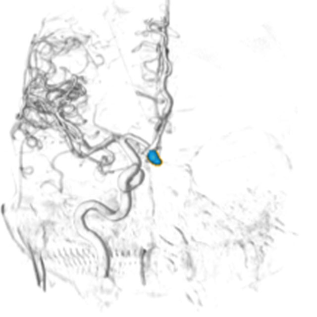
CADA Logo
CADA
Challenge User

Cerebral aneurysms are local dilations of arterial blood vessels caused by a weakness of the vessel wall. Subarachnoid hemorrhage (SAH) caused by the rupture of a cerebral aneurysm is a life-threatening condition associated with high mortality and morbidity. The mortality rate is above 40%, and even in case of survival cognitive impairment can affect patients for a long time. Major goals in image analysis are the detection and risk assessment of aneurysms. We, therefore, subdivided the challenge into three categories. The first task is finding the aneurysm; the second task is the accurate segmentation to allow for a longitudinal assessment of the development of suspicious aneurysms. The third task is the estimation of the rupture risk of the aneurysm.

SegPC-2021 Logo
SegPC-2021
Challenge User

This challenge is positioned towards robust segmentation of cells which is the first stage to build such a tool for plasma cell cancer, namely, Multiple Myeloma (MM), which is a type of blood cancer.

Carotid Artery Vessel Wall Segmentation Challenge
Challenge User

To segment the vessel wall of the carotid artery on black-blood MRI images

crossMoDA Logo
Cross-Modality Domain Adaptation Image Segmentation - 2021
Challenge User

The CrossMoDA challenge 2021 introduces the first large and multi-class medical dataset for unsupervised cross-modality Domain Adaptation.

FLARE Logo
FLARE21
Challenge User

Fast and Low GPU memory Abdominal oRgan sEgmentation Challenge

feta Logo
FeTA - Fetal Tissue Annotation Challenge
Challenge User

Fetal Tissue Annotation Challenge

AutoImplant2021 Logo
AutoImplant 2021
Challenge User

Second MICCAI Challenge on Automatic Cranial Implant Design (AutoImplant 2021)

DFU-2021 Logo
DFUC2021
Challenge User

Diabetic Foot Ulcer Challenge 2021

tiger Logo
TIGER
Challenge User

Grand challenge on automate assessment of tumor infiltrating lymphocytes in digital pathology slides of triple negative and Her2-positive breast cancers

PI-CAI Logo
The PI-CAI Challenge
Challenge User

Artificial Intelligence and Radiologists at Prostate Cancer Detection in MRI

AIROGS Logo
AIROGS
Challenge User

Artificial Intelligence for RObust Glaucoma Screening Challenge

CoNIC-Challenge Logo
CoNIC 2022
Challenge User

Colon Nuclei Identification and Counting Challenge 2022

EndoCV2022 Logo
EndoCV2022
Challenge User

Developing methods for "detection task'' and ''segmentation task'' for endoscopic video sequence data

MELA Logo
MELA2022
Challenge User

MICCAI 2022 MELA Challenge: A Large-Scale Detection Benchmark of 1,100 CT Scans for Mediastinal Lesion Analysis

Parse2022 Logo
Parse2022
Challenge User

It is of significant clinical interest to study pulmonary artery structures in the field of medical image analysis. One prerequisite step is to segment pulmonary artery structures from CT with high accuracy and low time-consuming. The segmentation of pulmonary artery structures benefits the quantification of its morphological changes for diagnosis of pulmonary hypertension and thoracic surgery. However, due to the complexity of pulmonary artery topology, automated segmentation of pulmonary artery topology is a challenging task. Besides, the open accessible large-scale CT data with well labeled pulmonary artery are scarce (The large variations of the topological structures from different patients make the annotation an extremely challenging process). The lack of well labeled pulmonary artery hinders the development of automatic pulmonary artery segmentation algorithm. Hence, we try to host the first Pulmonary ARtery SEgmentation challenge in MICCAI 2022 (Named Parse2022) to start a new research topic.

TDSC-ABUS2023 Logo
TDSC-ABUS2023
Challenge User

Tumor Detection, Segmentation and Classification Challenge on Automated 3D Breast Ultrasound

instance Logo
INSTANCE2022
Challenge User

The 2022 Intracranial Hemorrhage Segmentation Challenge on Non-Contrast head CT (NCCT)

BCNB Logo
BCNB
Challenge User

Early Breast Cancer Core-Needle Biopsy WSI Dataset

3DTeethSeg Logo
3D Teeth Scan Segmentation and Labeling Challenge MICCAI2022
Challenge User

Computer-aided design (CAD) tools have become increasingly popular in modern dentistry for highly accurate treatment planning. In particular, in orthodontic CAD systems, advanced intraoral scanners (IOSs) are now widely used as they provide precise digital surface models of the dentition. Such models can dramatically help dentists simulate teeth extraction, move, deletion, and rearrangement and therefore ease the prediction of treatment outcomes. Although IOSs are becoming widespread in clinical dental practice, there are only few contributions on teeth segmentation/labeling available in the literature and no publicly available database. A fundamental issue that appears with IOS data is the ability to reliably segment and identify teeth in scanned observations. Teeth segmentation and labelling is difficult as a result of the inherent similarities between teeth shapes as well as their ambiguous positions on jaws.

FLARE22 Logo
MICCAI FLARE 2022
Challenge User

MICCAI 2022 Fast and Low-resource semi-supervised Abdominal oRgan sEgmentation (FLARE) Challenge

AGGC22 Logo
AGGC22
Challenge User

ACROBAT Logo
ACROBAT 2023
Challenge User

The ACROBAT challenge aims to advance the development of WSI registration algorithms that can align WSIs of IHC-stained breast cancer tissue sections to corresponding tissue regions that were stained with H&E. All WSIs originate from routine diagnostic workflows.

SurgT Logo
SurgT: Surgical Tracking
Challenge User

This challenge consists of surgical videos with a target bounding box and the participants are expected to develop visual tracking algorithms to estimate the trajectory of the bounding box throughout the video-sequence.

DRAC22 Logo
Diabetic Retinopathy Analysis Challenge MICCAI2022
Challenge User

Diabetic Retinopathy (DR) lesions segmentation, image quality assessment and classification of proliferatived DR (PDR) and non-PDR in ultra-wide optical coherence tomography angiography mosaic (UW-OCTA-M) images

AMOS22 Logo
Multi-Modality Abdominal Multi-Organ Segmentation Challenge 2022
Challenge User

vessel-wall-segmentation-2022 Logo
Carotid Vessel Wall Segmentation and Atherosclerosis Diagnosis
Challenge User

ATM22 Logo
Multi-site, Multi-Domain Airway Tree Modeling (ATM’22)
Challenge User

Airway segmentation is a crucial step for the analysis of pulmonary diseases including asthma, bronchiectasis, and emphysema. The accurate segmentation based on X-Ray computed tomography (CT) enables the quantitative measurements of airway dimensions and wall thickness, which can reveal the abnormality of patients with chronic obstructive pulmonary disease (COPD). Besides, the extraction of patient-specific airway models from CT images is required for navigatiisted surgery.

PS-FH-AOP-2023 Logo
FH-PS-AOP challenge
Challenge User

Fetal Head and Pubic Symphysis Segmentation Challenge

isles22 Logo
Ischemic Stroke Lesion Segmentation Challenge
Challenge User

bci Logo
Breast Cancer Immunohistochemical Image Generation Challenge
Challenge User

The Breast Cancer Immunohistochemical Image Generation Challenge aims to directly generate IHC-stained breast cancer histopathology images from HE-stained images.

2023PAIP Logo
PAIP 2023: TC prediction in pancreatic and colon cancer
Challenge User

Tumor cellularity prediction in pancreatic cancer (supervised learning) and colon cancer (transfer learning)

UltrasoundEnhance2023 Logo
Ultrasound Image Enhancement challenge 2023
Challenge User

SegRap2023 Logo
SegRap 2023
Challenge User

A segmentation challenge with 200 patients (two modalities of CT images, 45 OARs and 2 GTVs).

ULS23 Logo
Universal Lesion Segmentation Challenge '23
Challenge User

JustRAIGS Logo
Justified Referral in AI Glaucoma Screening
Challenge User

PENGWIN Logo
Pelvic Bone Fragments with Injuries Segmentation Challenge
Challenge User

AIROGS Baseline Logo
AIROGS Baseline
Algorithm User

A baseline algorithm for the AIROGS challenge

SPIDER Baseline IIS Logo
SPIDER Baseline IIS
Algorithm User