L JJ
LJJ_0328
- China
- yes
- guangdong university
Statistics
- Member for 4 years, 5 months
- 1 challenge submissions
Activity Overview
Parse2022
Challenge UserIt is of significant clinical interest to study pulmonary artery structures in the field of medical image analysis. One prerequisite step is to segment pulmonary artery structures from CT with high accuracy and low time-consuming. The segmentation of pulmonary artery structures benefits the quantification of its morphological changes for diagnosis of pulmonary hypertension and thoracic surgery. However, due to the complexity of pulmonary artery topology, automated segmentation of pulmonary artery topology is a challenging task. Besides, the open accessible large-scale CT data with well labeled pulmonary artery are scarce (The large variations of the topological structures from different patients make the annotation an extremely challenging process). The lack of well labeled pulmonary artery hinders the development of automatic pulmonary artery segmentation algorithm. Hence, we try to host the first Pulmonary ARtery SEgmentation challenge in MICCAI 2022 (Named Parse2022) to start a new research topic.
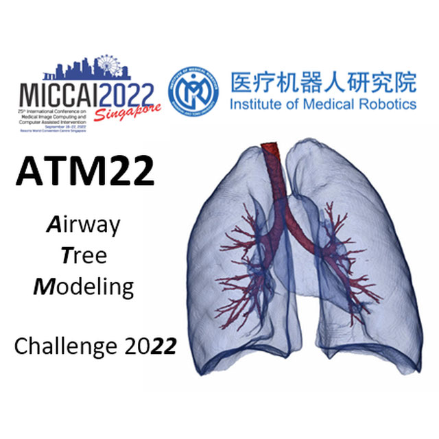
Multi-site, Multi-Domain Airway Tree Modeling (ATM’22)
Challenge UserAirway segmentation is a crucial step for the analysis of pulmonary diseases including asthma, bronchiectasis, and emphysema. The accurate segmentation based on X-Ray computed tomography (CT) enables the quantitative measurements of airway dimensions and wall thickness, which can reveal the abnormality of patients with chronic obstructive pulmonary disease (COPD). Besides, the extraction of patient-specific airway models from CT images is required for navigatiisted surgery.

