ming lei
sky2019
- China
- 西北工业大学
- 计算机学院
Statistics
- Member for 4 years, 5 months
Activity Overview
BreastPathQ: Cancer Cellularity Challenge 2019
Challenge UserSPIE-AAPM-NCI BreastPathQ:Cancer Circularity Challenge 2019: Participants will be tasked to develop an automated method for analyzing histology patches extracted from whole slide images and assign a score reflecting cancer cellularity for tumor burden assessment in each.

Gleason2019
Challenge UserMICCAI 2019 Automatic Prostate Gleason Grading Challenge: This challenge aims at the automatic Gleason grading of prostate cancer from H&E-stained histopathology images. This task is of critical importance because Gleason score is a strong prognostic predictor. On the other hand, it is very challenging because of the large degree of heterogeneity in the cellular and glandular patterns associated with each Gleason grade, leading to significant inter-observer variability, even among expert pathologists.

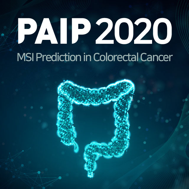
PAIP2020
Challenge UserBuilt on the success of its predecessor, PAIP2020 is the second challenge organized by the Pathology AI Platform (PAIP) and the Seoul National University Hospital (SNUH). PAIP2020 will proceed to not only detect whole tumor areas in colorectal cancers but also to classify their molecular subtypes, which will lead to characterization of their heterogeneity with respect to prognoses and therapeutic responses. All participants should predict one of the molecular carcinogenesis pathways, i.e., microsatellite instability(MSI) in colorectal cancer, by performing digital image analysis without clinical tests. This task has a high clinical relevance as the currently used procedure requires an extensive microscopic assessment by pathologists. Therefore, those automated algorithms would reduce the workload of pathologists as a diagnostic assistance.
Breast Cancer Segmentation
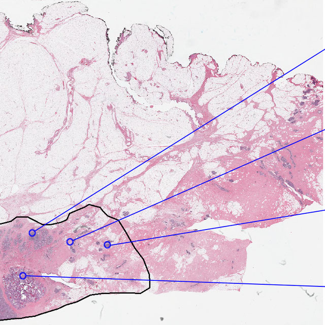
Challenge UserSemantic segmentation of histologic regions in scanned FFPE H&E stained slides of triple-negative breast cancer from The Cancer Genome Atlas. See: Amgad M, Elfandy H, ..., Gutman DA, Cooper LAD. Structured crowdsourcing enables convolutional segmentation of histology images. Bioinformatics. 2019. doi: 10.1093/bioinformatics/btz083

WSSS4LUAD
Challenge UserThe WSSS4LUAD dataset contains over 10,000 patches of lung adenocarcinoma from whole slide images from Guangdong Provincial People's Hospital and TCGA with image-level annotations. The goal of this challenge is to perform semantic segmentation for differentiating three important types of tissues in the WSIs of lung adenocarcinoma, including cancerous epithelial region, cancerous stroma region and normal region. Paticipants have to use image-level annotations to give pixel-level prediction.
3D Teeth Scan Segmentation and Labeling Challenge MICCAI2022
Challenge UserComputer-aided design (CAD) tools have become increasingly popular in modern dentistry for highly accurate treatment planning. In particular, in orthodontic CAD systems, advanced intraoral scanners (IOSs) are now widely used as they provide precise digital surface models of the dentition. Such models can dramatically help dentists simulate teeth extraction, move, deletion, and rearrangement and therefore ease the prediction of treatment outcomes. Although IOSs are becoming widespread in clinical dental practice, there are only few contributions on teeth segmentation/labeling available in the literature and no publicly available database. A fundamental issue that appears with IOS data is the ability to reliably segment and identify teeth in scanned observations. Teeth segmentation and labelling is difficult as a result of the inherent similarities between teeth shapes as well as their ambiguous positions on jaws.