Your mugshot

yanzhou su

syz

  •  China
  •  UESTC
  •  school of information and communication engineering
Statistics
  • Member for 4 years, 1 month
  • 35 challenge submissions

Activity Overview

VESSEL12 Logo
VESSEL12
Challenge User

The VESSEL12 challenge compares methods for automatic (and semi-automatic) segmentation of blood vessels in the lungs from CT images.

CRASS Logo
CRASS
Challenge User

CRASS stands for Chest Radiograph Anatomical Structure Segmentation. The challenge currently invites participants to send in results for clavicle segmentation algorithms.

CAUSE07 Logo
CAUSE07
Challenge User

The goal of CAUSE07 is to compare different algorithms to segment the caudate nucleaus from brain MRI scans.

PROMISE12 Logo
PROMISE12
Challenge User

The goal of this challenge is to compare interactive and (semi)-automatic segmentation algorithms for MRI of the prostate.

LUNA16 Logo
LUNA16
Challenge User

The LUNA16 challenge: automatic nodule detection on chest CT

CATARACTS Logo
CATARACTS
Challenge User

The Challenge on Automatic Tool Annotation for cataRACT Surgery aims at evaluating image-based tool detection algorithms in the context of the most common surgical procedure in the world.

SLIVER07 Logo
SLIVER07
Challenge User

The goal of this competition is to compare different algorithms to segment the liver from clinical 3D computed tomography (CT) scans.

IDRiD Logo
IDRiD
Challenge User

This challenge evaluates automated techniques for analysis of fundus photographs. We target segmentation of retinal lesions like exudates, microaneurysms, and hemorrhages and detection of the optic disc and fovea. Also, we seek grading of fundus images according to the severity level of DR and DME.

drive Logo
DRIVE
Challenge User

Develop a system to automatically segment vessels in human retina fundus images.

REFUGE Logo
REFUGE
Challenge User

The goal of the Retinal Fundus Glaucoma Challenge (REFUGE) is to evaluate and compare automated algorithms for glaucoma detection and optic disc/cup segmentation on a common dataset of retinal fundus images.

ANHIR Logo
ANHIR
Challenge User

The challenge focuses on comparing the accuracy (using manually annotated landmarks) and the approximate speed of automatic non-linear registration methods for aligning microscopy images of multi-stained histology tissue samples.

BreastPathQ Logo
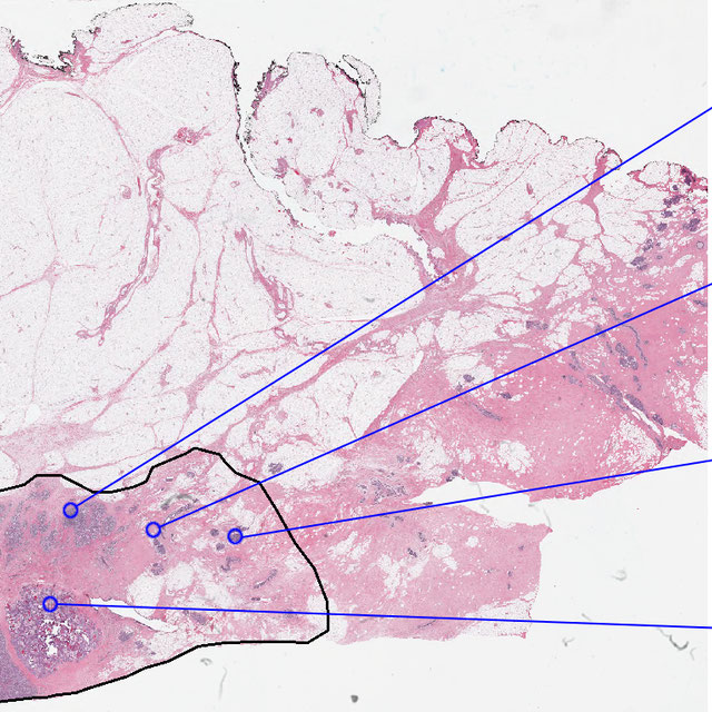
BreastPathQ: Cancer Cellularity Challenge 2019
Challenge User

SPIE-AAPM-NCI BreastPathQ:Cancer Circularity Challenge 2019: Participants will be tasked to develop an automated method for analyzing histology patches extracted from whole slide images and assign a score reflecting cancer cellularity for tumor burden assessment in each.
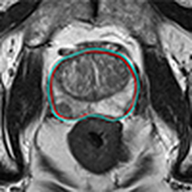
EAD2019 Logo
EAD2019
Challenge User

Endoscopic Artefact Detection (EAD) is a core problem and needed for realising robust computer-assisted tools. The EAD challenge has 3 tasks: 1) Multi-class artefact detection, 2) Region segmentation, 3) Detection generalisation.

iChallenges Logo
iChallenges
Challenge User

We organized a serial of challenges on different eye image modalities, such as REFUGE, PALM, RETOUCH, etc.

EndoVisSub2019-SCARED Logo
Stereo Correspondence and Reconstruction of Endoscopic Data
Challenge User

Stereo Correspondence and Reconstruction of Endoscopic Data

Gleason2019 Logo
Gleason2019
Challenge User

MICCAI 2019 Automatic Prostate Gleason Grading Challenge: This challenge aims at the automatic Gleason grading of prostate cancer from H&E-stained histopathology images. This task is of critical importance because Gleason score is a strong prognostic predictor. On the other hand, it is very challenging because of the large degree of heterogeneity in the cellular and glandular patterns associated with each Gleason grade, leading to significant inter-observer variability, even among expert pathologists.
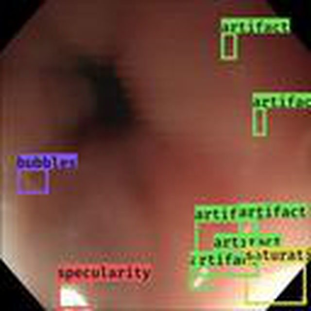
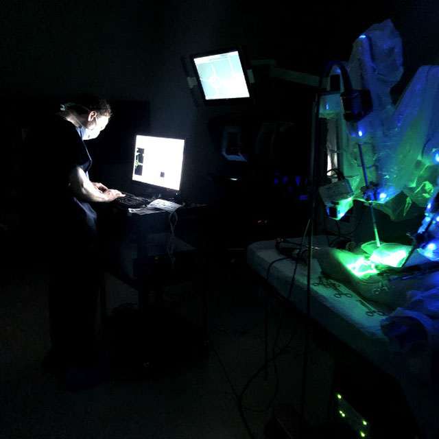
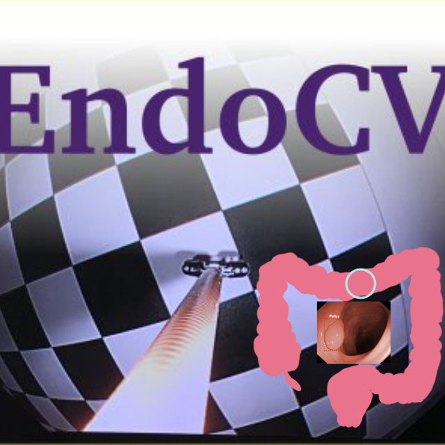
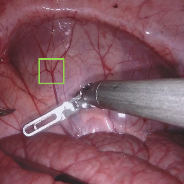
DigestPath2019 Logo
DigestPath2019
Challenge User

Welcome to Digestive-System Pathological Detection and Segmentation Challenge 2019. This competition is part of the MICCAI 2019 Challenge.

odir2019 Logo
ODIR-2019
Challenge User

北京大学国际眼底图像智能识别竞赛 Peking University International Competition on Ocular Disease Intelligent Recognition

LYSTO Logo
Lymphocyte Assessment Hackathon
Challenge User

Lymphocyte Assessment Hackathon in conjunction with the MICCAI COMPAY 2019 Workshop on Computational Pathology

ECDP2020 Logo
HEROHE
Challenge User

Unlike previous challenges, this proposes to find an image analysis algorithm to identify HER2-positive from HER2-negative breast cancer specimens evaluating only the morphological features present on the HE slide, without the staining patterns of IHC.

LNDb Logo
LNDb Challenge
Challenge User

Lung cancer screening and Fleischner follow-up determination in chest CT through nodule detection, segmentation and characterization

RibFrac Logo
RibFrac
Challenge User

Rib Fracture Detection and Classification Challenge: A large-scale benchmark of 660 CT scans with ~5,000 rib fractures (around 80Gb)

SARAS-ESAD
Challenge User

This challenge is part of Medical Imaging with Deep Learning conference, 2020. The conference is held between 6 ‑ 8 July 2020 in Montréal. The SARAS (Smart Autonomous Robotic Assistant Surgeon) EU consortium, www.saras-project.eu, is working towards replacing the assistant surgeon in MIS with two assistive robotic arms. To accomplish that, an artificial intelligence based system is required which not only can understand the complete surgical scene but also detect the actions being performed by the main surgeon. This information can later be used infer the response required from the autonomous assistant surgeon.

AutoImplant Logo
AutoImplant
Challenge User

the MICCAI 2020 Cranial Implant Design Challenge

qubiq Logo
QUBIQ
Challenge User

Quantification of Uncertainties in Biomedical Image Segmentation Challenge

RIADD Logo
RIADD (ISBI-2021)
Challenge User

Retinal Image Analysis for multi-Disease Detection

MitoEM Logo
MitoEM
Challenge User

Large-scale 3D mitochondria instance segmentation benchmark

covid-segmentation Logo
COVID-19 LUNG CT LESION SEGMENTATION CHALLENGE - 2020
Challenge User

This challenge will create the platform to evaluate emerging methods for the segmentation and quantification of lung lesions caused by SARS-CoV-2 infection from CT images.

VALDO Logo
Where is VALDO?
Challenge User

Vascular Lesion Detection Challenge at MICCAI 2021

SegPC-2021 Logo
SegPC-2021
Challenge User

This challenge is positioned towards robust segmentation of cells which is the first stage to build such a tool for plasma cell cancer, namely, Multiple Myeloma (MM), which is a type of blood cancer.

EndoCV2021 Logo
EndoCV2021
Challenge User

Endoscopy Computer Vision Challenge 2021

Carotid Artery Vessel Wall Segmentation Challenge
Challenge User

To segment the vessel wall of the carotid artery on black-blood MRI images

crossMoDA Logo
Cross-Modality Domain Adaptation Image Segmentation - 2021
Challenge User

The CrossMoDA challenge 2021 introduces the first large and multi-class medical dataset for unsupervised cross-modality Domain Adaptation.

BrainPTM-2021 Logo
BrainPTM 2021
Challenge User

Brain Pre-surgical Tractography Mapping (BrainPTM) in real clinical scans.

FLARE Logo
FLARE21
Challenge User

Fast and Low GPU memory Abdominal oRgan sEgmentation Challenge

feta Logo
FeTA - Fetal Tissue Annotation Challenge
Challenge User

Fetal Tissue Annotation Challenge

AutoImplant2021 Logo
AutoImplant 2021
Challenge User

Second MICCAI Challenge on Automatic Cranial Implant Design (AutoImplant 2021)

DFU-2021 Logo
DFUC2021
Challenge User

Diabetic Foot Ulcer Challenge 2021

QUBIQ21 Logo
QUBIQ2021
Challenge User

Quantification of Uncertainties in Biomedical Image Segmentation Challenge 2021

tiger Logo
TIGER
Challenge User

Grand challenge on automate assessment of tumor infiltrating lymphocytes in digital pathology slides of triple negative and Her2-positive breast cancers

PI-CAI Logo
The PI-CAI Challenge
Challenge User

Artificial Intelligence and Radiologists at Prostate Cancer Detection in MRI

ultra-low-dose-PET Logo
Ultra-low Dose PET Imaging Challenge
Challenge User

This challenge aims to develop deep learning algorithms capable of recovering high-quality imaging from low-dose scans on this ultra-low-dose PET scanner.

CoNIC-Challenge Logo
CoNIC 2022
Challenge User

Colon Nuclei Identification and Counting Challenge 2022

kipa22 Logo
KiPA22 (Regular Challenge)
Challenge User

The challenge is aimed to segment kidney, renal tumors, arteries, and veins from computed tomography angiography (CTA) images in one inference.

TDSC-ABUS2023 Logo
TDSC-ABUS2023
Challenge User

Tumor Detection, Segmentation and Classification Challenge on Automated 3D Breast Ultrasound

instance Logo
INSTANCE2022
Challenge User

The 2022 Intracranial Hemorrhage Segmentation Challenge on Non-Contrast head CT (NCCT)

BCNB Logo
BCNB
Challenge User

Early Breast Cancer Core-Needle Biopsy WSI Dataset

ATLAS Logo
ATLAS R2.0 - Stroke Lesion Segmentation
Challenge User

Anatomical Tracings of Lesions After Stroke

SynthRAD2023 Logo
SynthRAD2023
Challenge User

SynthRAD is the first challenge on automatic generation of synthetic computed tomography (sCT) for radiotherapy

FLARE22 Logo
MICCAI FLARE 2022
Challenge User

MICCAI 2022 Fast and Low-resource semi-supervised Abdominal oRgan sEgmentation (FLARE) Challenge

AGGC22 Logo
AGGC22
Challenge User

autoPET Logo
autoPET
Challenge User

Automatic lesion segmentation in whole-body FDG-PET/CT

ACROBAT Logo
ACROBAT 2023
Challenge User

The ACROBAT challenge aims to advance the development of WSI registration algorithms that can align WSIs of IHC-stained breast cancer tissue sections to corresponding tissue regions that were stained with H&E. All WSIs originate from routine diagnostic workflows.

SurgT Logo
SurgT: Surgical Tracking
Challenge User

This challenge consists of surgical videos with a target bounding box and the participants are expected to develop visual tracking algorithms to estimate the trajectory of the bounding box throughout the video-sequence.

P2ILF Logo
Preoperative to Intraoperative Laparoscopy Fusion
Challenge User

Preoperative to Intraoperative Laparoscopy Fusion

DRAC22 Logo
Diabetic Retinopathy Analysis Challenge MICCAI2022
Challenge User

Diabetic Retinopathy (DR) lesions segmentation, image quality assessment and classification of proliferatived DR (PDR) and non-PDR in ultra-wide optical coherence tomography angiography mosaic (UW-OCTA-M) images

AMOS22 Logo
Multi-Modality Abdominal Multi-Organ Segmentation Challenge 2022
Challenge User

vessel-wall-segmentation-2022 Logo
Carotid Vessel Wall Segmentation and Atherosclerosis Diagnosis
Challenge User

crossmoda2022 Logo
Cross-Modality Domain Adaptation: Segmentation & Classification
Challenge User

The CrossMoDA 2022 challenge is the second edition of the first large and multi-class medical dataset for unsupervised cross-modality Domain Adaptation.

ATM22 Logo
Multi-site, Multi-Domain Airway Tree Modeling (ATM’22)
Challenge User

Airway segmentation is a crucial step for the analysis of pulmonary diseases including asthma, bronchiectasis, and emphysema. The accurate segmentation based on X-Ray computed tomography (CT) enables the quantitative measurements of airway dimensions and wall thickness, which can reveal the abnormality of patients with chronic obstructive pulmonary disease (COPD). Besides, the extraction of patient-specific airway models from CT images is required for navigatiisted surgery.

isles22 Logo
Ischemic Stroke Lesion Segmentation Challenge
Challenge User

NeurIPS22-CellSeg Logo
Cell Segmentation in Multi-modality Microscopy Images
Challenge User

Weakly Supervised Cell Segmentation in Multi-modality High-resolution Microscopy Images

2023PAIP Logo
PAIP 2023: TC prediction in pancreatic and colon cancer
Challenge User

Tumor cellularity prediction in pancreatic cancer (supervised learning) and colon cancer (transfer learning)

HaN-Seg2023 Logo
The Head and Neck Organ-at-Risk CT & MR Segmentation Challenge
Challenge User

A semantic multimodal segmentation challenge comprising 30 organs at risk

RnR-ExM Logo
Robust Non-rigid Registration Challenge for Expansion Microscopy
Challenge User

autoPET-II Logo
autoPET-II
Challenge User

Automated Lesion Segmentation in PET/CT - Domain Generalization

toothfairy Logo
ToothFairy: Cone-Beam Computed Tomography Segmentation Challenge
Challenge User

This is the first edition of the ToothFairy challenge organized by the University of Modena and Reggio Emilia with the collaboration of Raudboud University. This challenge aims at pushing the development of deep learning frameworks to segment the Inferior Alveolar Canal (IAC) by incrementally extending the amount of publicly available 3D-annotated Cone Beam Computed Tomography (CBCT) scans. CBCT modality is becoming increasingly important for treatment planning and diagnosis in implant dentistry and maxillofacial surgery. The three-dimensional information acquired with CBCT can be crucial to plan a vast number of surgical interventions with the aim of preserving noble anatomical structures such as the Inferior Alveolar Canal (IAC), which contains the homonymous nerve (Inferior Alveolar Nerve, IAN). Deep learning models can support medical personnel in surgical planning procedures by providing a voxel-level segmentation of the IAN automatically extracted from CBCT scans.
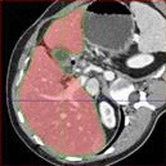
spider Logo
SPIDER
Challenge User

LNQ2023 Logo
LNQ2023
Challenge User

Accurate lymph node size estimation is critical for staging cancer patients, initial therapeutic management, and in longitudinal scans, assessing response to therapy. Current standard practice for quantifying lymph node size is based on a variety of criteria that use unidirectional or bidirectional measurements on just one or a few nodes, typically on just one axial slice. But humans have hundreds of lymph nodes, any number of which may be enlarged to various degrees due to disease or immune response. While a normal lymph node may be approximately 5mm in diameter, a diseased lymph node may be several cm in diameter. The mediastinum, the anatomical area between the lungs and around the heart, may contain ten or more lymph nodes, often with three or more enlarged greater than 1cm. Accurate segmentation in 3D would provide more information to evaluate lymph node disease.

MultiCenterAorta Logo
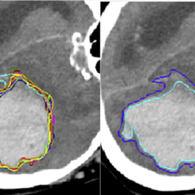
SEG.A. - Segmentation of the Aorta
Challenge User

Segmentation, modeling and visualization of the arterial tree are still a challenge in medical image analysis. The main track of this challenge deals with the fully automatic segmentation of the aortic vessel tree in computed tomography images. Optionally, teams can submit tailored solutions for meshing and visualization of the vessel tree.

SPPIN Logo
Surgical Planning in Pediatric Neuroblastoma
Challenge User

MedFM2023 Logo
Foundation Model Prompting for Medical Image Classification
Challenge User

The primary objective of this challenge is to promote the development and evaluation of model adaptation techniques for medical image classification to leverage the existing foundation models.

DENTEX Logo
DENTEX - MICCAI23
Challenge User

Dental Enumeration and Diagnosis on Panoramic X- rays Challenge

SegRap2023 Logo
SegRap 2023
Challenge User

A segmentation challenge with 200 patients (two modalities of CT images, 45 OARs and 2 GTVs).

THOMPSON-Challenge Logo
The Trauma THOMPSON Challenge
Challenge User

ULS23 Logo
Universal Lesion Segmentation Challenge '23
Challenge User