Your mugshot

suman chaudhary

chaudharysuman560

  •  China
  •  Taiyuan University of Technology
  •  Department of information and computer
  •  Website
Statistics
  • Member for 3 years, 7 months
  • 70 challenge submissions

Activity Overview

LUNA16 Logo
LUNA16
Challenge User

The LUNA16 challenge: automatic nodule detection on chest CT

Decathlon-10 Logo
Decathlon
Challenge User

The Medical Segmentation Decathlon challenge tests the generalisability of machine learning algorithms when applied to 10 different semantic segmentation task.

LNDb Logo
LNDb Challenge
Challenge User

Lung cancer screening and Fleischner follow-up determination in chest CT through nodule detection, segmentation and characterization

COVID-CT Logo
CT diagnosis of COVID-19
Challenge User

Coronavirus disease 2019 (COVID-19) has infected more than 1.3 million individuals all over the world and caused more than 106,000 deaths. One major hurdle in controlling the spreading of this disease is the inefficiency and shortage of medical tests. To mitigate the inefficiency and shortage of existing tests for COVID-19, we propose this competition to encourage the development of effective Deep Learning techniques to diagnose COVID-19 based on CT images. The problem we want to solve is to classify each CT image into positive COVID-19 (the image has clinical findings of COVID-19) or negative COVID-19 ( the image does not have clinical findings of COVID-19). It’s a binary classification problem based on CT images.

covid-segmentation Logo
COVID-19 LUNG CT LESION SEGMENTATION CHALLENGE - 2020
Challenge User

This challenge will create the platform to evaluate emerging methods for the segmentation and quantification of lung lesions caused by SARS-CoV-2 infection from CT images.

fastPET-LD Logo
fastPET-LD
Challenge User

The purpose of this challenge is the detection of “hot spots” in fast PET scan, that is locations that have an elevated standard uptake value (SUV) and potential clinical significance. Corresponding CT scans are also provided. The ground truth, common to both datasets, was generated by a nuclear medicine expert. It consists of a 3-D segmentation map of the hot spots as well as a text file containing the position and size of 3D cuboid bounding box for each hot spot.

NODE21 Logo
NODE21
Challenge User

NODE21: generate and detect nodules on chest radiographs

tiger Logo
TIGER
Challenge User

Grand challenge on automate assessment of tumor infiltrating lymphocytes in digital pathology slides of triple negative and Her2-positive breast cancers

STOIC2021 Logo
STOIC2021 - COVID-19 AI Challenge
Challenge User

COVID-19 Artificial Intelligence Challenge: automated diagnosis, and prognostic evaluation based on computed tomography

CXR-COVID19 Logo
Chest XR COVID-19 detection
Challenge User

Build AI models to detect COVID-19 using Chest X-ray images

PI-CAI Logo
The PI-CAI Challenge
Challenge User

Artificial Intelligence and Radiologists at Prostate Cancer Detection in MRI

AIROGS Logo
AIROGS
Challenge User

Artificial Intelligence for RObust Glaucoma Screening Challenge

CoNIC-Challenge Logo
CoNIC 2022
Challenge User

Colon Nuclei Identification and Counting Challenge 2022

EndoCV2022 Logo
EndoCV2022
Challenge User

Developing methods for "detection task'' and ''segmentation task'' for endoscopic video sequence data

MELA Logo
MELA2022
Challenge User

MICCAI 2022 MELA Challenge: A Large-Scale Detection Benchmark of 1,100 CT Scans for Mediastinal Lesion Analysis

kipa22 Logo
KiPA22 (Regular Challenge)
Challenge User

The challenge is aimed to segment kidney, renal tumors, arteries, and veins from computed tomography angiography (CTA) images in one inference.

Parse2022 Logo
Parse2022
Challenge User

It is of significant clinical interest to study pulmonary artery structures in the field of medical image analysis. One prerequisite step is to segment pulmonary artery structures from CT with high accuracy and low time-consuming. The segmentation of pulmonary artery structures benefits the quantification of its morphological changes for diagnosis of pulmonary hypertension and thoracic surgery. However, due to the complexity of pulmonary artery topology, automated segmentation of pulmonary artery topology is a challenging task. Besides, the open accessible large-scale CT data with well labeled pulmonary artery are scarce (The large variations of the topological structures from different patients make the annotation an extremely challenging process). The lack of well labeled pulmonary artery hinders the development of automatic pulmonary artery segmentation algorithm. Hence, we try to host the first Pulmonary ARtery SEgmentation challenge in MICCAI 2022 (Named Parse2022) to start a new research topic.

TDSC-ABUS2023 Logo
TDSC-ABUS2023
Challenge User

Tumor Detection, Segmentation and Classification Challenge on Automated 3D Breast Ultrasound

instance Logo
INSTANCE2022
Challenge User

The 2022 Intracranial Hemorrhage Segmentation Challenge on Non-Contrast head CT (NCCT)

BCNB Logo
BCNB
Challenge User

Early Breast Cancer Core-Needle Biopsy WSI Dataset

ATLAS Logo
ATLAS R2.0 - Stroke Lesion Segmentation
Challenge User

Anatomical Tracings of Lesions After Stroke

3DTeethSeg Logo
3D Teeth Scan Segmentation and Labeling Challenge MICCAI2022
Challenge User

Computer-aided design (CAD) tools have become increasingly popular in modern dentistry for highly accurate treatment planning. In particular, in orthodontic CAD systems, advanced intraoral scanners (IOSs) are now widely used as they provide precise digital surface models of the dentition. Such models can dramatically help dentists simulate teeth extraction, move, deletion, and rearrangement and therefore ease the prediction of treatment outcomes. Although IOSs are becoming widespread in clinical dental practice, there are only few contributions on teeth segmentation/labeling available in the literature and no publicly available database. A fundamental issue that appears with IOS data is the ability to reliably segment and identify teeth in scanned observations. Teeth segmentation and labelling is difficult as a result of the inherent similarities between teeth shapes as well as their ambiguous positions on jaws.
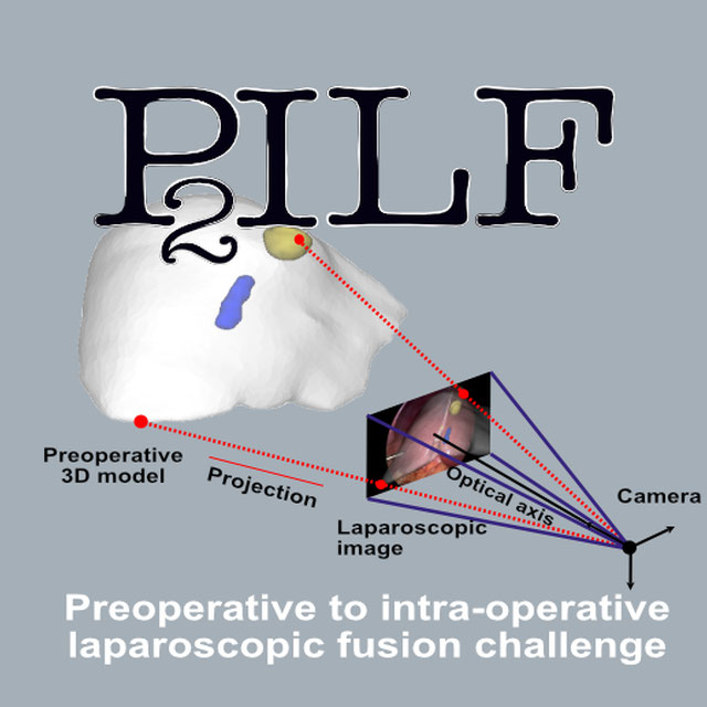
P2ILF Logo
Preoperative to Intraoperative Laparoscopy Fusion
Challenge User

Preoperative to Intraoperative Laparoscopy Fusion

K2S Logo
K2S: from undersampled K-space To automatic Segmentation
Challenge User

K2S: from undersampled K-space To automatic Segmentation

DRAC22 Logo
Diabetic Retinopathy Analysis Challenge MICCAI2022
Challenge User

Diabetic Retinopathy (DR) lesions segmentation, image quality assessment and classification of proliferatived DR (PDR) and non-PDR in ultra-wide optical coherence tomography angiography mosaic (UW-OCTA-M) images

AMOS22 Logo
Multi-Modality Abdominal Multi-Organ Segmentation Challenge 2022
Challenge User

curious2022 Logo
Brain shift with Intraoperative Ultrasound - Segmentation tasks
Challenge User

MEGC2022 Logo
ACMMM MEGC2022: Facial Micro-Expression Grand Challenge
Challenge User

Spotting Facial Macro- and Micro-Expressions in Long Videos

isles22 Logo
Ischemic Stroke Lesion Segmentation Challenge
Challenge User

MedFM2023 Logo
Foundation Model Prompting for Medical Image Classification
Challenge User

The primary objective of this challenge is to promote the development and evaluation of model adaptation techniques for medical image classification to leverage the existing foundation models.