Your mugshot

Ashwini Kumar Pal

ashwinids

  •  India
  •  Not Associated with an Institution
  •  Not Associated with an Institution
Statistics
  • Member for 3 years, 6 months
  • 1 challenge submissions

Activity Overview

RIADD Logo
RIADD (ISBI-2021)
Challenge User

Retinal Image Analysis for multi-Disease Detection

covid-segmentation Logo
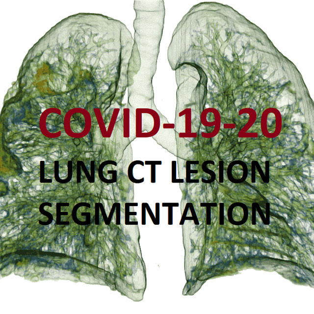
COVID-19 LUNG CT LESION SEGMENTATION CHALLENGE - 2020
Challenge User

This challenge will create the platform to evaluate emerging methods for the segmentation and quantification of lung lesions caused by SARS-CoV-2 infection from CT images.

VALDO Logo
Where is VALDO?
Challenge User

Vascular Lesion Detection Challenge at MICCAI 2021

SegPC-2021 Logo
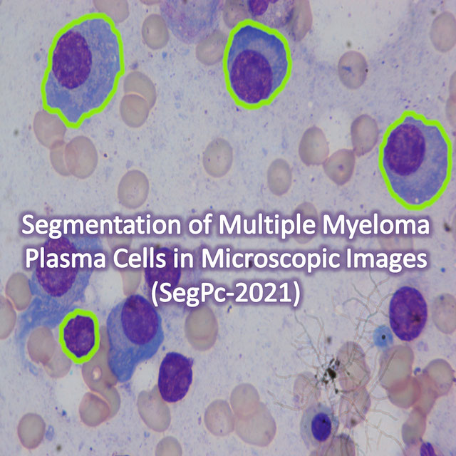
SegPC-2021
Challenge User

This challenge is positioned towards robust segmentation of cells which is the first stage to build such a tool for plasma cell cancer, namely, Multiple Myeloma (MM), which is a type of blood cancer.

EndoCV2021 Logo
EndoCV2021
Challenge User

Endoscopy Computer Vision Challenge 2021

FUSC Logo
Foot Ulcer Segmentation Challenge
Challenge User

BrainPTM-2021 Logo
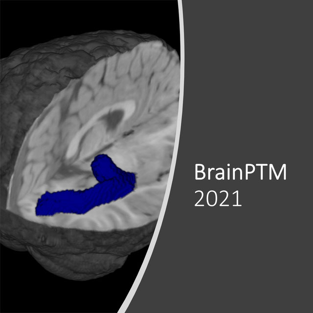
BrainPTM 2021
Challenge User

Brain Pre-surgical Tractography Mapping (BrainPTM) in real clinical scans.

CholecTriplet2021 Logo
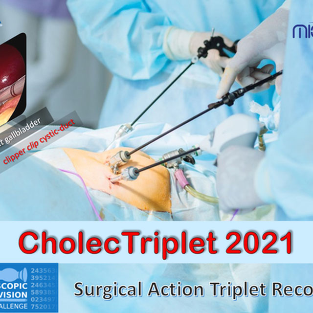
CholecTriplet 2021
Challenge User

EndoVis Sub-challenge for Surgical Action Triplet Recognition

PAIP2021 Logo
PAIP2021
Challenge User

PAIP 2021 Challenge; Perineural invasion in multiple organ cancer (colon, prostate and pancreatobiliary tract)

FLARE Logo
FLARE21
Challenge User

Fast and Low GPU memory Abdominal oRgan sEgmentation Challenge

NuCLS Logo
NuCLS
Challenge User

Classification, Localization and Segmentation of nuclei in scanned FFPE H&E stained slides of triple-negative breast cancer from The Cancer Genome Atlas. See: Amgad et al. 2021. arXiv:2102.09099 [cs.CV].

BCSegmentation Logo
Breast Cancer Segmentation
Challenge User

Semantic segmentation of histologic regions in scanned FFPE H&E stained slides of triple-negative breast cancer from The Cancer Genome Atlas. See: Amgad M, Elfandy H, ..., Gutman DA, Cooper LAD. Structured crowdsourcing enables convolutional segmentation of histology images. Bioinformatics. 2019. doi: 10.1093/bioinformatics/btz083

feta Logo
FeTA - Fetal Tissue Annotation Challenge
Challenge User

Fetal Tissue Annotation Challenge

AutoImplant2021 Logo
AutoImplant 2021
Challenge User

Second MICCAI Challenge on Automatic Cranial Implant Design (AutoImplant 2021)

SARAS-MESAD Logo
SARAS-MESAD
Challenge User

This challenge is organized under MICCAI 2021, the 24th International Conference on Medical Image Computing and Computer Assisted Intervention. The event will be held from September 27th to October 1st 2021 in Strasbourg, France. The challenge focuses on multi-domain surgeon action detection.

NODE21 Logo
NODE21
Challenge User

NODE21: generate and detect nodules on chest radiographs

MIDOG2021 Logo
MIDOG Challenge 2021
Challenge User

Mitosis Domain Generalization Challenge 2021 (part of MICCAI 2021)

tiger Logo
TIGER
Challenge User

Grand challenge on automate assessment of tumor infiltrating lymphocytes in digital pathology slides of triple negative and Her2-positive breast cancers

STOIC2021 Logo
STOIC2021 - COVID-19 AI Challenge
Challenge User

COVID-19 Artificial Intelligence Challenge: automated diagnosis, and prognostic evaluation based on computed tomography

CXR-COVID19 Logo
Chest XR COVID-19 detection
Challenge User

Build AI models to detect COVID-19 using Chest X-ray images

PI-CAI Logo
The PI-CAI Challenge
Challenge User

Artificial Intelligence and Radiologists at Prostate Cancer Detection in MRI

ultra-low-dose-PET Logo
Ultra-low Dose PET Imaging Challenge
Challenge User

This challenge aims to develop deep learning algorithms capable of recovering high-quality imaging from low-dose scans on this ultra-low-dose PET scanner.

AIROGS Logo
AIROGS
Challenge User

Artificial Intelligence for RObust Glaucoma Screening Challenge

CoNIC-Challenge Logo
CoNIC 2022
Challenge User

Colon Nuclei Identification and Counting Challenge 2022

cholectriplet2022 Logo
CholecTriplet2022: Surgical Action Triplet Detection
Challenge User

Surgical Action Triplet Detection 2022

EndoCV2022 Logo
EndoCV2022
Challenge User

Developing methods for "detection task'' and ''segmentation task'' for endoscopic video sequence data

MELA Logo
MELA2022
Challenge User

MICCAI 2022 MELA Challenge: A Large-Scale Detection Benchmark of 1,100 CT Scans for Mediastinal Lesion Analysis

Parse2022 Logo
Parse2022
Challenge User

It is of significant clinical interest to study pulmonary artery structures in the field of medical image analysis. One prerequisite step is to segment pulmonary artery structures from CT with high accuracy and low time-consuming. The segmentation of pulmonary artery structures benefits the quantification of its morphological changes for diagnosis of pulmonary hypertension and thoracic surgery. However, due to the complexity of pulmonary artery topology, automated segmentation of pulmonary artery topology is a challenging task. Besides, the open accessible large-scale CT data with well labeled pulmonary artery are scarce (The large variations of the topological structures from different patients make the annotation an extremely challenging process). The lack of well labeled pulmonary artery hinders the development of automatic pulmonary artery segmentation algorithm. Hence, we try to host the first Pulmonary ARtery SEgmentation challenge in MICCAI 2022 (Named Parse2022) to start a new research topic.

TDSC-ABUS2023 Logo
TDSC-ABUS2023
Challenge User

Tumor Detection, Segmentation and Classification Challenge on Automated 3D Breast Ultrasound

instance Logo
INSTANCE2022
Challenge User

The 2022 Intracranial Hemorrhage Segmentation Challenge on Non-Contrast head CT (NCCT)

RAVIR Logo
RAVIR
Challenge User

A dataset for semantic segmentation and quantitative analysis of retinal arteries and veins in infrared reflectance imaging

DFUC2022 Logo
DFUC 2022
Challenge User

Diabetes is a global epidemic affecting around 425 million people and expected to rise to 629 million by 2045. Diabetic Foot Ulcer (DFU) is a severe condition that can result from the disease. The rise of the condition over the last decades is a challenge for healthcare systems. Cases of DFU usually lead to severe conditions that greatly prolongs treatment and result in limb amputation or death. Recent research focuses on creating detection algorithms to monitor their condition to improve patient care and reduce strain on healthcare systems. Work between Manchester Metropolitan University, Lancashire Teaching Hospitals and Manchester University NHS Foundation Trust has created an international repository of up to 11,000 DFU images. Analysis of ulcer regions is a key for DFU management. Delineation of ulcers is time-consuming. With effort from the lead scientists of the UK, US, India and New Zealand, this challenge promotes novel work in DFU segmentation and promote interdisciplinary researcher collaboration.

ATLAS Logo
ATLAS R2.0 - Stroke Lesion Segmentation
Challenge User

Anatomical Tracings of Lesions After Stroke

SynthRAD2023 Logo
SynthRAD2023
Challenge User

SynthRAD is the first challenge on automatic generation of synthetic computed tomography (sCT) for radiotherapy

3DTeethSeg Logo
3D Teeth Scan Segmentation and Labeling Challenge MICCAI2022
Challenge User

Computer-aided design (CAD) tools have become increasingly popular in modern dentistry for highly accurate treatment planning. In particular, in orthodontic CAD systems, advanced intraoral scanners (IOSs) are now widely used as they provide precise digital surface models of the dentition. Such models can dramatically help dentists simulate teeth extraction, move, deletion, and rearrangement and therefore ease the prediction of treatment outcomes. Although IOSs are becoming widespread in clinical dental practice, there are only few contributions on teeth segmentation/labeling available in the literature and no publicly available database. A fundamental issue that appears with IOS data is the ability to reliably segment and identify teeth in scanned observations. Teeth segmentation and labelling is difficult as a result of the inherent similarities between teeth shapes as well as their ambiguous positions on jaws.

FLARE22 Logo
MICCAI FLARE 2022
Challenge User

MICCAI 2022 Fast and Low-resource semi-supervised Abdominal oRgan sEgmentation (FLARE) Challenge

AGGC22 Logo
AGGC22
Challenge User

autoPET Logo
autoPET
Challenge User

Automatic lesion segmentation in whole-body FDG-PET/CT

ACROBAT Logo
ACROBAT 2023
Challenge User

The ACROBAT challenge aims to advance the development of WSI registration algorithms that can align WSIs of IHC-stained breast cancer tissue sections to corresponding tissue regions that were stained with H&E. All WSIs originate from routine diagnostic workflows.

SurgT Logo
SurgT: Surgical Tracking
Challenge User

This challenge consists of surgical videos with a target bounding box and the participants are expected to develop visual tracking algorithms to estimate the trajectory of the bounding box throughout the video-sequence.

SLCN Logo
Surface Learning for Clinical Neuroimaging
Challenge User

Prediction of developmental phenotypes (age at birth and cognitive test scores) from cortical surface imaging data

P2ILF Logo
Preoperative to Intraoperative Laparoscopy Fusion
Challenge User

Preoperative to Intraoperative Laparoscopy Fusion

K2S Logo
K2S: from undersampled K-space To automatic Segmentation
Challenge User

K2S: from undersampled K-space To automatic Segmentation

DRAC22 Logo
Diabetic Retinopathy Analysis Challenge MICCAI2022
Challenge User

Diabetic Retinopathy (DR) lesions segmentation, image quality assessment and classification of proliferatived DR (PDR) and non-PDR in ultra-wide optical coherence tomography angiography mosaic (UW-OCTA-M) images

AMOS22 Logo
Multi-Modality Abdominal Multi-Organ Segmentation Challenge 2022
Challenge User

curious2022 Logo
Brain shift with Intraoperative Ultrasound - Segmentation tasks
Challenge User

vessel-wall-segmentation-2022 Logo
Carotid Vessel Wall Segmentation and Atherosclerosis Diagnosis
Challenge User

crossmoda2022 Logo
Cross-Modality Domain Adaptation: Segmentation & Classification
Challenge User

The CrossMoDA 2022 challenge is the second edition of the first large and multi-class medical dataset for unsupervised cross-modality Domain Adaptation.

ATM22 Logo
Multi-site, Multi-Domain Airway Tree Modeling (ATM’22)
Challenge User

Airway segmentation is a crucial step for the analysis of pulmonary diseases including asthma, bronchiectasis, and emphysema. The accurate segmentation based on X-Ray computed tomography (CT) enables the quantitative measurements of airway dimensions and wall thickness, which can reveal the abnormality of patients with chronic obstructive pulmonary disease (COPD). Besides, the extraction of patient-specific airway models from CT images is required for navigatiisted surgery.

PS-FH-AOP-2023 Logo
FH-PS-AOP challenge
Challenge User

Fetal Head and Pubic Symphysis Segmentation Challenge

MIDOG2022 Logo
MItosis DOmain Generalization Challenge 2022
Challenge User

isles22 Logo
Ischemic Stroke Lesion Segmentation Challenge
Challenge User

NeurIPS22-CellSeg Logo
Cell Segmentation in Multi-modality Microscopy Images
Challenge User

Weakly Supervised Cell Segmentation in Multi-modality High-resolution Microscopy Images

bci Logo
Breast Cancer Immunohistochemical Image Generation Challenge
Challenge User

The Breast Cancer Immunohistochemical Image Generation Challenge aims to directly generate IHC-stained breast cancer histopathology images from HE-stained images.

AUTO-RTP Logo
Fully Automated Radiotherapy Treatment Planning Challenge
Challenge User

Evaluating fully automated external beam radiotherapy treatment planning in prostate cancer

2023PAIP Logo
PAIP 2023: TC prediction in pancreatic and colon cancer
Challenge User

Tumor cellularity prediction in pancreatic cancer (supervised learning) and colon cancer (transfer learning)

SNEMI3D Logo
SNEMI3D: 3D Segmentation of neurites in EM images
Challenge User

The challenge is organized in the context of the IEEE International Symposium on Biomedical Imaging, 2013. The old evaluation site (http://brainiac2.snemi3d.org/SNEMI3D/) will be replaced by this one.

HaN-Seg2023 Logo
The Head and Neck Organ-at-Risk CT & MR Segmentation Challenge
Challenge User

A semantic multimodal segmentation challenge comprising 30 organs at risk

ENDO-AID Logo
Endometrial Carcinoma Detection in Pipelle biopsies
Challenge User

Evaluation platform as reference benchmark for algorithms that can predict endometrial carcinoma on whole-slide images of Pipelle sampled endometrial slides stained in H&E, based on the test data set used in our project.

RnR-ExM Logo
Robust Non-rigid Registration Challenge for Expansion Microscopy
Challenge User

XPRESS Logo
Xray Projectomic Reconstruction Extracting Segment with Skeleton
Challenge User

autoPET-II Logo
autoPET-II
Challenge User

Automated Lesion Segmentation in PET/CT - Domain Generalization

toothfairy Logo
ToothFairy: Cone-Beam Computed Tomography Segmentation Challenge
Challenge User

This is the first edition of the ToothFairy challenge organized by the University of Modena and Reggio Emilia with the collaboration of Raudboud University. This challenge aims at pushing the development of deep learning frameworks to segment the Inferior Alveolar Canal (IAC) by incrementally extending the amount of publicly available 3D-annotated Cone Beam Computed Tomography (CBCT) scans. CBCT modality is becoming increasingly important for treatment planning and diagnosis in implant dentistry and maxillofacial surgery. The three-dimensional information acquired with CBCT can be crucial to plan a vast number of surgical interventions with the aim of preserving noble anatomical structures such as the Inferior Alveolar Canal (IAC), which contains the homonymous nerve (Inferior Alveolar Nerve, IAN). Deep learning models can support medical personnel in surgical planning procedures by providing a voxel-level segmentation of the IAN automatically extracted from CBCT scans.

LNQ2023 Logo
LNQ2023
Challenge User

Accurate lymph node size estimation is critical for staging cancer patients, initial therapeutic management, and in longitudinal scans, assessing response to therapy. Current standard practice for quantifying lymph node size is based on a variety of criteria that use unidirectional or bidirectional measurements on just one or a few nodes, typically on just one axial slice. But humans have hundreds of lymph nodes, any number of which may be enlarged to various degrees due to disease or immune response. While a normal lymph node may be approximately 5mm in diameter, a diseased lymph node may be several cm in diameter. The mediastinum, the anatomical area between the lungs and around the heart, may contain ten or more lymph nodes, often with three or more enlarged greater than 1cm. Accurate segmentation in 3D would provide more information to evaluate lymph node disease.

ARCADE Logo
ARCADE-MICCAI2023
Challenge User

UltrasoundEnhance2023 Logo
Ultrasound Image Enhancement challenge 2023
Challenge User

MultiCenterAorta Logo
SEG.A. - Segmentation of the Aorta
Challenge User

Segmentation, modeling and visualization of the arterial tree are still a challenge in medical image analysis. The main track of this challenge deals with the fully automatic segmentation of the aortic vessel tree in computed tomography images. Optionally, teams can submit tailored solutions for meshing and visualization of the vessel tree.

SPPIN Logo
Surgical Planning in Pediatric Neuroblastoma
Challenge User

MedFM2023 Logo
Foundation Model Prompting for Medical Image Classification
Challenge User

The primary objective of this challenge is to promote the development and evaluation of model adaptation techniques for medical image classification to leverage the existing foundation models.

DENTEX Logo
DENTEX - MICCAI23
Challenge User

Dental Enumeration and Diagnosis on Panoramic X- rays Challenge

TopCoW23 Logo
Topology-Aware Anatomical Segmentation of the Circle of Willis
Challenge User

Segment the Circle of Willis (CoW) vessel components for both CTA and MRA

SegRap2023 Logo
SegRap 2023
Challenge User

A segmentation challenge with 200 patients (two modalities of CT images, 45 OARs and 2 GTVs).

LDCTIQAC2023 Logo
Low-dose Computed Tomography Perceptual Image Quality Assessment
Challenge User

CL-Detection2023 Logo
CL-Detection 2023
Challenge User

Cephalometric landmark detection in lateral x-ray images

surgtoolloc23 Logo
Endoscopic surgical tool localization using tool presence labels
Challenge User

OCELOT2023 Logo
OCELOT 2023: Cell Detection from Cell-Tissue Interaction
Challenge User

THOMPSON-Challenge Logo
The Trauma THOMPSON Challenge
Challenge User

BONBID-HIE2023 Logo
Hypoxic Ischemic Encephalopathy Lesion Segmentation Challenge
Challenge User

ULS23 Logo
Universal Lesion Segmentation Challenge '23
Challenge User

JustRAIGS Logo
Justified Referral in AI Glaucoma Screening
Challenge User

lightmycells Logo
Light My Cells : Bright Field to Fluorescence Imaging Challenge
Challenge User

Join the Light My Cells France-Bioimaging challenge! Enhance biology and microscopy by contributing to the development of new image-to-image deep labelling methods. The task: predict the best-focused output images of several fluorescently labelled organelles from label-free transmitted light input images. Dive into the future of imaging with us! 🌐🔬 #LightMyCellsChallenge

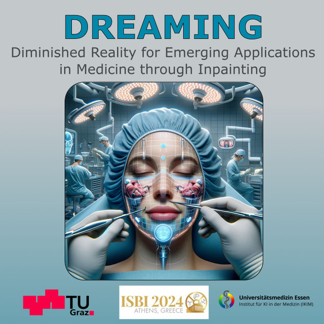
DREAMING Logo
Diminished Reality for Emerging Applications in Medicine
Challenge User

The Diminished Reality for Emerging Applications in Medicine through Inpainting (DREAMING) challenge seeks to pioneer the integration of Diminished Reality (DR) into oral and maxillofacial surgery. While Augmented Reality (AR) has been extensively explored in medicine, DR remains largely uncharted territory. DR involves virtually removing real objects from the environment by replacing them with their background. Recent inpainting methods present an opportunity for real-time DR applications without scene knowledge. DREAMING focuses on implementing such methods to fill obscured regions in surgery scenes with realistic backgrounds, emphasizing the complex facial anatomy and patient diversity. The challenge provides a dataset of synthetic yet photorealistic surgery scenes featuring humans, simulating an operating room setting. Participants are tasked with developing algorithms that seamlessly remove disruptions caused by medical instruments and hands, offering surgeons an unimpeded view of the operative site.

LEOPARD Logo
The LEOPARD Challenge
Challenge User