Tomas Iesmantas
Tomas
- Lithuania
- Kaunas University of Technology
- Department of Mathematics and Natural Sciences
Statistics
- Member for 3 years
- 9 challenge submissions
Activity Overview

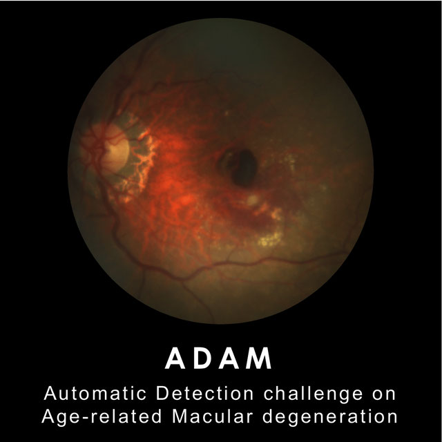
IDRiD
Challenge UserThis challenge evaluates automated techniques for analysis of fundus photographs. We target segmentation of retinal lesions like exudates, microaneurysms, and hemorrhages and detection of the optic disc and fovea. Also, we seek grading of fundus images according to the severity level of DR and DME.


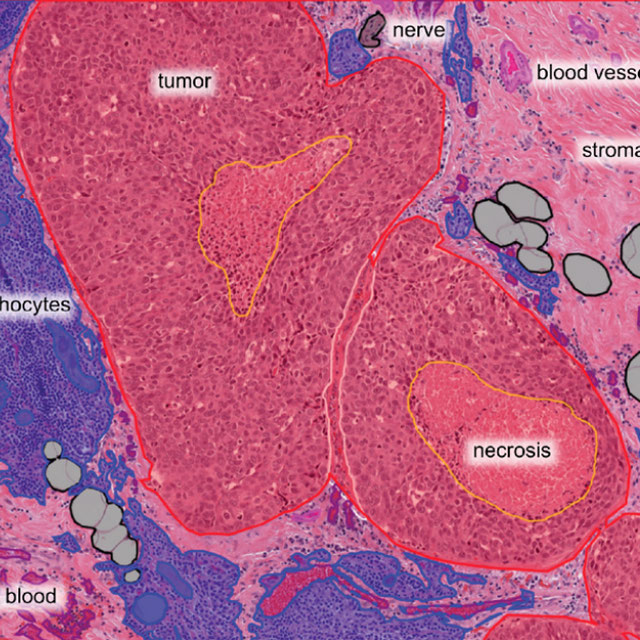
Breast Cancer Segmentation
Challenge UserSemantic segmentation of histologic regions in scanned FFPE H&E stained slides of triple-negative breast cancer from The Cancer Genome Atlas. See: Amgad M, Elfandy H, ..., Gutman DA, Cooper LAD. Structured crowdsourcing enables convolutional segmentation of histology images. Bioinformatics. 2019. doi: 10.1093/bioinformatics/btz083
Parse2022
Challenge UserIt is of significant clinical interest to study pulmonary artery structures in the field of medical image analysis. One prerequisite step is to segment pulmonary artery structures from CT with high accuracy and low time-consuming. The segmentation of pulmonary artery structures benefits the quantification of its morphological changes for diagnosis of pulmonary hypertension and thoracic surgery. However, due to the complexity of pulmonary artery topology, automated segmentation of pulmonary artery topology is a challenging task. Besides, the open accessible large-scale CT data with well labeled pulmonary artery are scarce (The large variations of the topological structures from different patients make the annotation an extremely challenging process). The lack of well labeled pulmonary artery hinders the development of automatic pulmonary artery segmentation algorithm. Hence, we try to host the first Pulmonary ARtery SEgmentation challenge in MICCAI 2022 (Named Parse2022) to start a new research topic.


LNQ2023
Challenge UserAccurate lymph node size estimation is critical for staging cancer patients, initial therapeutic management, and in longitudinal scans, assessing response to therapy. Current standard practice for quantifying lymph node size is based on a variety of criteria that use unidirectional or bidirectional measurements on just one or a few nodes, typically on just one axial slice. But humans have hundreds of lymph nodes, any number of which may be enlarged to various degrees due to disease or immune response. While a normal lymph node may be approximately 5mm in diameter, a diseased lymph node may be several cm in diameter. The mediastinum, the anatomical area between the lungs and around the heart, may contain ten or more lymph nodes, often with three or more enlarged greater than 1cm. Accurate segmentation in 3D would provide more information to evaluate lymph node disease.