Yiyu Hong
Arontier_HYY
- South Korea
- Arontier
- Research
Statistics
- Member for 5 years, 3 months
- 66 challenge submissions
Activity Overview

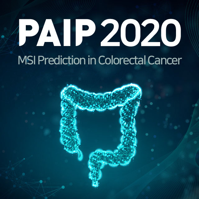
PAIP2020
Challenge UserBuilt on the success of its predecessor, PAIP2020 is the second challenge organized by the Pathology AI Platform (PAIP) and the Seoul National University Hospital (SNUH). PAIP2020 will proceed to not only detect whole tumor areas in colorectal cancers but also to classify their molecular subtypes, which will lead to characterization of their heterogeneity with respect to prognoses and therapeutic responses. All participants should predict one of the molecular carcinogenesis pathways, i.e., microsatellite instability(MSI) in colorectal cancer, by performing digital image analysis without clinical tests. This task has a high clinical relevance as the currently used procedure requires an extensive microscopic assessment by pathologists. Therefore, those automated algorithms would reduce the workload of pathologists as a diagnostic assistance.
Breast Cancer Segmentation
Challenge UserSemantic segmentation of histologic regions in scanned FFPE H&E stained slides of triple-negative breast cancer from The Cancer Genome Atlas. See: Amgad M, Elfandy H, ..., Gutman DA, Cooper LAD. Structured crowdsourcing enables convolutional segmentation of histology images. Bioinformatics. 2019. doi: 10.1093/bioinformatics/btz083

WSSS4LUAD
Challenge UserThe WSSS4LUAD dataset contains over 10,000 patches of lung adenocarcinoma from whole slide images from Guangdong Provincial People's Hospital and TCGA with image-level annotations. The goal of this challenge is to perform semantic segmentation for differentiating three important types of tissues in the WSIs of lung adenocarcinoma, including cancerous epithelial region, cancerous stroma region and normal region. Paticipants have to use image-level annotations to give pixel-level prediction.
SEG.A. - Segmentation of the Aorta
Challenge UserSegmentation, modeling and visualization of the arterial tree are still a challenge in medical image analysis. The main track of this challenge deals with the fully automatic segmentation of the aortic vessel tree in computed tomography images. Optionally, teams can submit tailored solutions for meshing and visualization of the vessel tree.