Your mugshot

xiangde luo

xiangdeluo

  •  China
  •  University of Electronic Science and Technology of China
  •  School of Mechanical and Electrical Engineering
  •  Website
Statistics
  • Member for 5 years, 1 month
  • 24 challenge submissions

Activity Overview

VESSEL12 Logo
VESSEL12
Challenge User

The VESSEL12 challenge compares methods for automatic (and semi-automatic) segmentation of blood vessels in the lungs from CT images.

CRASS Logo
CRASS
Challenge User

CRASS stands for Chest Radiograph Anatomical Structure Segmentation. The challenge currently invites participants to send in results for clavicle segmentation algorithms.

PROMISE12 Logo
PROMISE12
Challenge User

The goal of this challenge is to compare interactive and (semi)-automatic segmentation algorithms for MRI of the prostate.

IDRiD Logo
IDRiD
Challenge User

This challenge evaluates automated techniques for analysis of fundus photographs. We target segmentation of retinal lesions like exudates, microaneurysms, and hemorrhages and detection of the optic disc and fovea. Also, we seek grading of fundus images according to the severity level of DR and DME.

drive Logo
DRIVE
Challenge User

Develop a system to automatically segment vessels in human retina fundus images.

REFUGE Logo
REFUGE
Challenge User

The goal of the Retinal Fundus Glaucoma Challenge (REFUGE) is to evaluate and compare automated algorithms for glaucoma detection and optic disc/cup segmentation on a common dataset of retinal fundus images.

PROSTATEx Logo
PROSTATEx
Challenge User

Classification of clinical significance of prostate lesions using multi-parametric MRI data

HC18 Logo
HC18
Challenge User

Automated measurement of fetal head circumference using 2D ultrasound images

CHAOS Logo
CHAOS
Challenge User

In this challenge, you segment the liver in CT data, and segment liver, spleen, and kidneys in MRI data.

Decathlon-10 Logo
Decathlon
Challenge User

The Medical Segmentation Decathlon challenge tests the generalisability of machine learning algorithms when applied to 10 different semantic segmentation task.

KiTS19 Logo
KiTS19
Challenge User

2019 Kidney and Kidney Tumor Segmentation Challenge

PAIP2019 Logo
PAIP 2019
Challenge User

PAIP2019: Liver Cancer Segmentation Task 1: Liver Cancer Segmentation Task 2: Viable Tumor Burden Estimation

VerSe2019 Logo
VerSe`19
Challenge User

Vertebrae labelling and segmentation on a spine dataset on an unprecedented 150 CT scans with voxel-level vertebral annotations.

AGE Logo
AGE
Challenge User

ANGLE CLOSURE GLAUCOMA EVALUATION CHALLENGE

AMD Logo
iChallenge-AMD
Challenge User

Age-related Macular Degeneration Challenge

StructSeg2019 Logo
StructSeg2019
Challenge User

Welcome to Automatic Structure Segmentation for Radiotherapy Planning Challenge 2019. This competition is part of the MICCAI 2019 Challenge.

odir2019 Logo
ODIR-2019
Challenge User

北京大学国际眼底图像智能识别竞赛 Peking University International Competition on Ocular Disease Intelligent Recognition

ECDP2020 Logo
HEROHE
Challenge User

Unlike previous challenges, this proposes to find an image analysis algorithm to identify HER2-positive from HER2-negative breast cancer specimens evaluating only the morphological features present on the HE slide, without the staining patterns of IHC.

TN-SCUI2020 Logo
Thyroid Nodule Segmentation and Classification
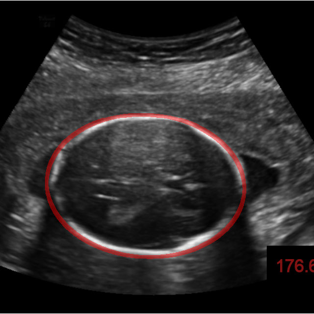
Challenge User

The main topic of this TN-SCUI2020 challenge is finding automatic algorithms to accurately classify the thyroid nodules in ultrasound images. It will provide the biggest public dataset of thyroid nodule with over 4500 patient cases from different ages, genders, and were collected using different ultrasound machines. Each ultrasound image is provided with its ground truth class (benign or maglinant) and a detailed delineation of the nodule. This challenge will provide a unique opportunity for participants from different backgrounds (e.g. academia, industry, and government, etc.) to compare their algorithms in an impartial way.
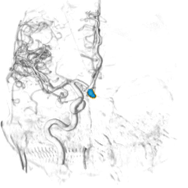
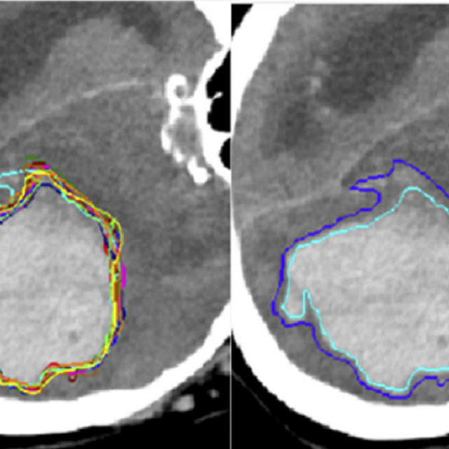
CADA Logo
CADA
Challenge User

Cerebral aneurysms are local dilations of arterial blood vessels caused by a weakness of the vessel wall. Subarachnoid hemorrhage (SAH) caused by the rupture of a cerebral aneurysm is a life-threatening condition associated with high mortality and morbidity. The mortality rate is above 40%, and even in case of survival cognitive impairment can affect patients for a long time. Major goals in image analysis are the detection and risk assessment of aneurysms. We, therefore, subdivided the challenge into three categories. The first task is finding the aneurysm; the second task is the accurate segmentation to allow for a longitudinal assessment of the development of suspicious aneurysms. The third task is the estimation of the rupture risk of the aneurysm.

qubiq Logo
QUBIQ
Challenge User

Quantification of Uncertainties in Biomedical Image Segmentation Challenge

MitoEM Logo
MitoEM
Challenge User

Large-scale 3D mitochondria instance segmentation benchmark

covid-segmentation Logo
COVID-19 LUNG CT LESION SEGMENTATION CHALLENGE - 2020
Challenge User

This challenge will create the platform to evaluate emerging methods for the segmentation and quantification of lung lesions caused by SARS-CoV-2 infection from CT images.

SegPC-2021 Logo
SegPC-2021
Challenge User

This challenge is positioned towards robust segmentation of cells which is the first stage to build such a tool for plasma cell cancer, namely, Multiple Myeloma (MM), which is a type of blood cancer.

EndoCV2021 Logo
EndoCV2021
Challenge User

Endoscopy Computer Vision Challenge 2021

crossMoDA Logo
Cross-Modality Domain Adaptation Image Segmentation - 2021
Challenge User

The CrossMoDA challenge 2021 introduces the first large and multi-class medical dataset for unsupervised cross-modality Domain Adaptation.

WSSS4LUAD Logo
WSSS4LUAD
Challenge User

The WSSS4LUAD dataset contains over 10,000 patches of lung adenocarcinoma from whole slide images from Guangdong Provincial People's Hospital and TCGA with image-level annotations. The goal of this challenge is to perform semantic segmentation for differentiating three important types of tissues in the WSIs of lung adenocarcinoma, including cancerous epithelial region, cancerous stroma region and normal region. Paticipants have to use image-level annotations to give pixel-level prediction.

instance Logo
INSTANCE2022
Challenge User

The 2022 Intracranial Hemorrhage Segmentation Challenge on Non-Contrast head CT (NCCT)

AMOS22 Logo
Multi-Modality Abdominal Multi-Organ Segmentation Challenge 2022
Challenge User

LNQ2023 Logo
LNQ2023
Challenge User

Accurate lymph node size estimation is critical for staging cancer patients, initial therapeutic management, and in longitudinal scans, assessing response to therapy. Current standard practice for quantifying lymph node size is based on a variety of criteria that use unidirectional or bidirectional measurements on just one or a few nodes, typically on just one axial slice. But humans have hundreds of lymph nodes, any number of which may be enlarged to various degrees due to disease or immune response. While a normal lymph node may be approximately 5mm in diameter, a diseased lymph node may be several cm in diameter. The mediastinum, the anatomical area between the lungs and around the heart, may contain ten or more lymph nodes, often with three or more enlarged greater than 1cm. Accurate segmentation in 3D would provide more information to evaluate lymph node disease.

MedFM2023 Logo
Foundation Model Prompting for Medical Image Classification
Challenge User

The primary objective of this challenge is to promote the development and evaluation of model adaptation techniques for medical image classification to leverage the existing foundation models.

SegRap2023 Logo
SegRap 2023
Challenge Editor

A segmentation challenge with 200 patients (two modalities of CT images, 45 OARs and 2 GTVs).

HookNet-TLS Logo
HookNet-TLS
Algorithm User

A model for detection of TLS and GC in histopathology.