Summer Newsletter 2026
Published 13 July 2026
Welcome to the Summer Edition of Grand Challenge Insights!
In this blog, we will share the latest updates and highlights from our Grand Challenge community.
💶 About our funding
A question we sometimes get: how does Grand Challenge sustain itself?
Like most research infrastructure, it isn't free to run: hosting, storage, and compute at our scale come with real costs. Part of that is covered by fees from users of the platform, but the majority is covered by research grants brought in by our own research group at Radboudumc.
That combination, user contributions plus grant funding, is what allows Grand Challenge to keep operating as it always has: as a non-profit platform run by Radboudumc for the medical imaging research community. Of course, we're also open to long-term partnerships with other academic institutions, other research groups, and other organisations that want to run challenges on the platform and help us jointly develop and maintain it.
Contact us at support@grand-challenge.org to discuss your interest!
🛠 New features of Grand Challenge
Below you will find some highlights of the new features on our platform. For an extensive list, refer to the Cycle updates published at the end of every cycle.
If you have feature requests or suggestions for improvements, you can contact us at support@grand-challenge.org.
🖊 Annotations & Viewer
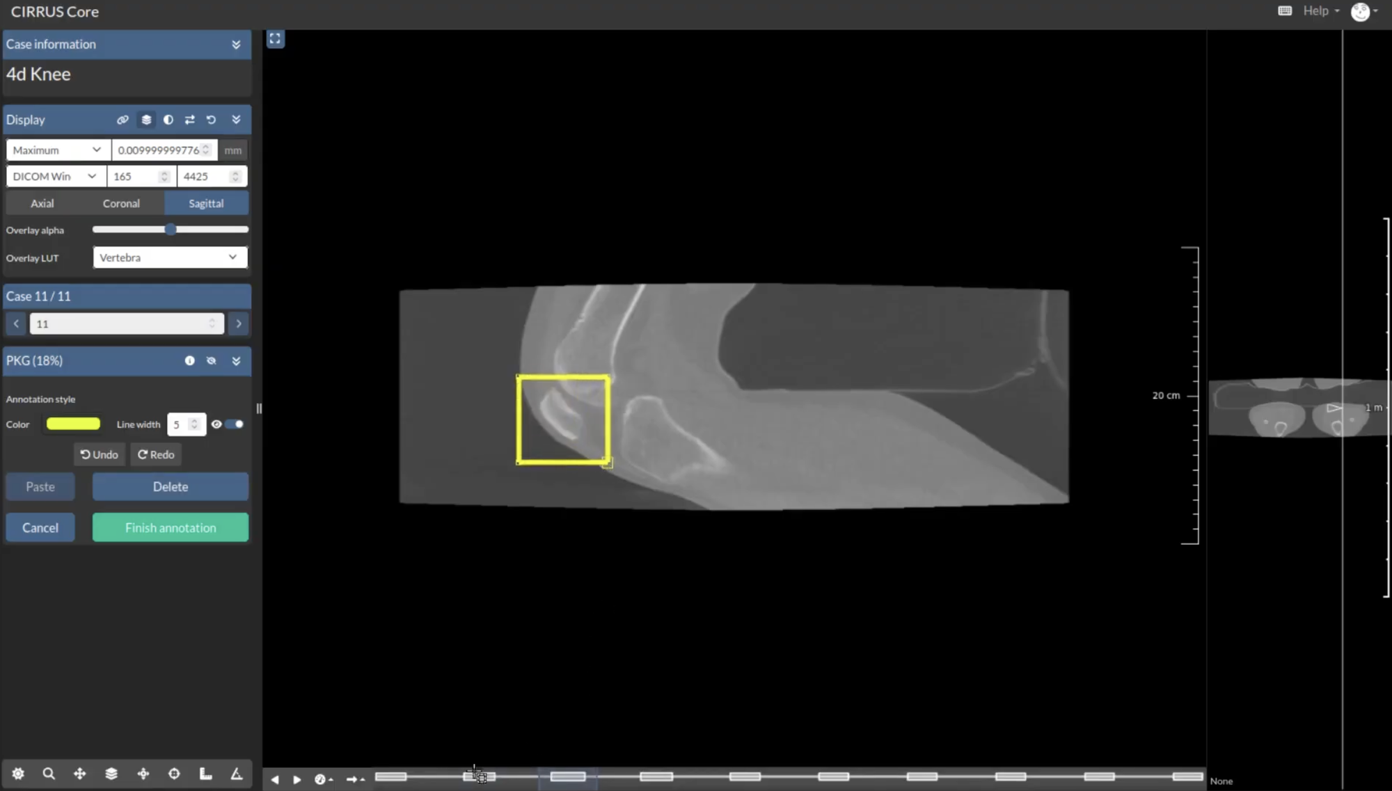
4D Annotations
It is now possible to create polygonal annotations on 4D time series images that include a time component, recording exactly which time frame an annotation was made on. This mode activates automatically when a time series
image is uploaded to a reader study, and the resulting annotations carry an extra time index field. Most
annotation types are supported, with the exception of points, two-line angles, and masks.
Platform & Viewer Improvements
Two useful improvements were made to the platform and the viewer. A new Your Work section in the main
dropdown gives quick access to all challenges, reader studies, archives, and algorithms, to help find what you
need quickly. On top of that, the 3D viewer now automatically selects the best viewing plane for any annotation
type, so there is no longer a need to manually reorient the view every time an annotation is clicked.
Scaling Annotations
A proof of concept was built this cycle to support loading over a million annotations in the client-side
pathology viewer. A WebGL approach makes it possible to render annotations in real time, with interactive
highlighting and label display. Bidirectional highlighting is also in place, meaning selecting an annotation
in the sidebar highlights it in the viewer and vice versa. Currently, the proof of concept covers point
annotations for algorithm jobs and archive items.
View-Plane Metadata on Annotations
Annotations now remember the orientation they were made in. Reviewers no longer have to guess: a real
help for non-cardinal orientations like those in ultrasound. Clicking an annotation now centres it and restores
the original orientation and zoom, and the field can also be set from algorithms.
🏆 Challenges & Evaluations
Renewed Evaluations and Submissions Overview
The submissions and evaluations pages have been redesigned to make debugging a lot easier. Now, error messages
come with clear explanations and suggested next steps, so participants and admins can quickly understand what
went wrong and how to fix it. The submission list also includes direct links to detailed evaluation pages where
all relevant information is easy to find.
Available Budget of Challenges
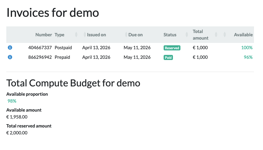
Challenge organisers can now see their budget consumption in near real time, rather than only getting a warning
when it runs low. The admin page has been renamed Invoice & Budget, giving a clear picture of what has
been spent so far.
Serverless Asynchronous Tasks
Background tasks like image validation, container imports, and job scheduling have moved to a serverless AWS
setup, replacing the less reliable Celery system. It is a behind-the-scenes change, but it lowers latency and
makes the platform steadier to use.
📖 Reader Studies
GPU-based Interactive Algorithms in Reader Studies
Interactive algorithms in reader studies can now use GPU processing, running through the new invocations
endpoints so the startup cost is paid once and every invocation after that is fast. CIRRUS uploads the input,
invokes the algorithm, and shows the result as an overlay. Connecting one to a question is currently manual,
so contact support if you would like to try it.
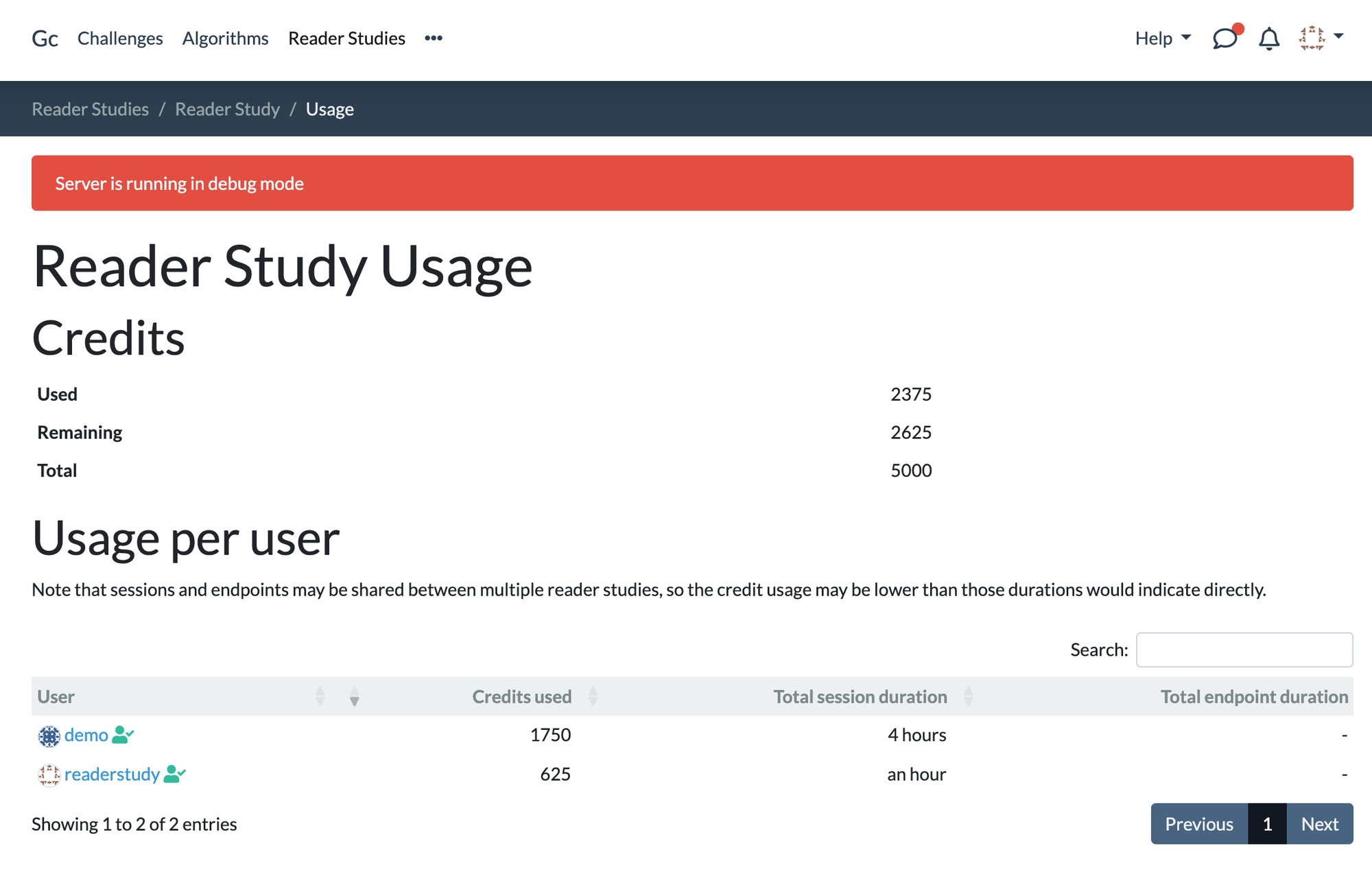
Budget for Reader Studies
Reviewing a reader study no longer depends on having budget available. Editors can always launch a study in
read-only mode to review their readers' answers, and a new overview shows how much budget has been used, how
much is left, and how it breaks down per reader.
Algorithm Endpoints & Invocations
Algorithm Endpoints continued to take shape over the last cycles. We have implemented the backend changes to launch Algorithm Endpoints, which host algorithm containers on GPU-enabled infrastructure and allow an algorithm to be invoked multiple times.
We also completed the invocation model and added REST API methods for requesting and querying invocations, letting CIRRUS and external applications invoke an endpoint and retrieve its outputs.
💡 Developer Blog posts
"April 2026 Cycle Report"
Read all about the platform improvements from our RSE team in April. Algorithm endpoints progress, "Your Work" search shortcut, million-annotation viewer prototype, 4D timed annotations.
"May 2026 Cycle Report"
Read all about the platform improvements from our RSE team in May. Clearer submission debugging, algorithm endpoint invocation groundwork, consistent 3D plane matching, CIRRUS bug fixes.
"June 2026 Cycle Report"
Read all about the platform improvements from our RSE team in June. Real-time budget tracking, GPU-powered interactive algorithms, serverless task migration, view-plane metadata for annotations.
🔦 Highlighted Challenges

🥅 Goal: MAMA-SYNTH is a challenge focused on synthesizing virtual post-contrast breast MRI from pre-contrast
T1-weighted MRI, with the aim of supporting the development of contrast-reduced and contrast-free breast MRI
workflows. By enabling rigorous comparison of virtual contrast-enhancement methods, the challenge addresses key concerns around patient safety, environmental impact, and accessibility of advanced imaging.
✍🏼 Task: Single-timepoint 2D peak-enhancement post-contrast breast DCE-MRI slice synthesis from pre-contrast
T1-weighted MRI inputs.
🥅 Goal: autoPET-V is a challenge focused on interactive, clinician-in-the-loop lesion segmentation in whole-body PET/CT. The challenge aims to support adaptive and clinically meaningful human–AI collaboration by evaluating how accurately, robustly, and efficiently algorithms improve lesion segmentation when given sparse corrective input. It addresses key challenges in automated tumor lesion segmentation, including domain shifts across tracers, scanners, and centres, physiological uptake false positives; and difficult low-contrast cases.
✍🏼 Task: Interactive lesion segmentation in whole-body PET/CT. Algorithms generate an initial segmentation of tumor lesions and iteratively refine it using sparse corrective scribbles targeting false-positive and false-negative regions.
🥅 Goal: HECKTOR 2026 is a challenge focused on head and neck tumor lesion segmentation, TN staging, and prognosis prediction. The challenge aims to support an end-to-end clinical imaging pipeline for treatment planning, improved risk stratification, and more accurate outcome prediction in head and neck cancer workflows.
✍🏼 Task: Unified end-to-end head and neck oncology pipeline covering tumor segmentation, TN staging classification, and prognosis prediction. The segmentation task delineates primary tumor and nodal tumor regions, while the downstream tasks predict TN stage and recurrence-free survival.
🔦 Highlighted Algorithms
isvisible
This algorithm detects and segments intestinal spirochetosis (IS) in H&E stained colon whole-slide images. It runs automatically on a single slide and outputs a full-slide binary segmentation mask at x20 magnification, identifying tissue regions that could potentially indicate the presence of spirochetosis. The model is based on a DeepLabV3Plus architecture with a Mix Transformer encoder, trained on 289 colonic biopsy whole-slide images from Radboudumc.