Bram van Ginneken
BramVanGinneken
- Netherlands
- Radboud University Medical Center
- Medical Imaging
- Website
Organizations
Statistics
- Member for 11 years, 5 months
- 2470 algorithms run
Activity Overview


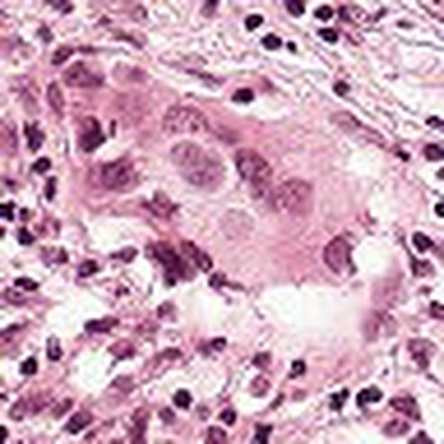

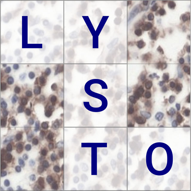

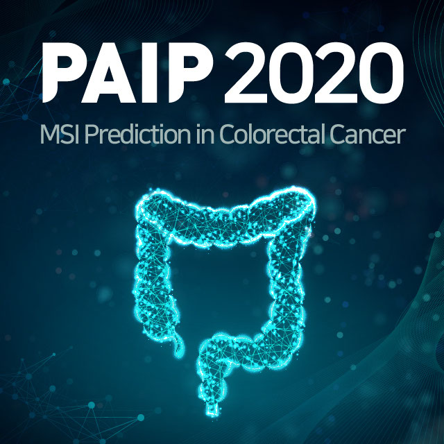
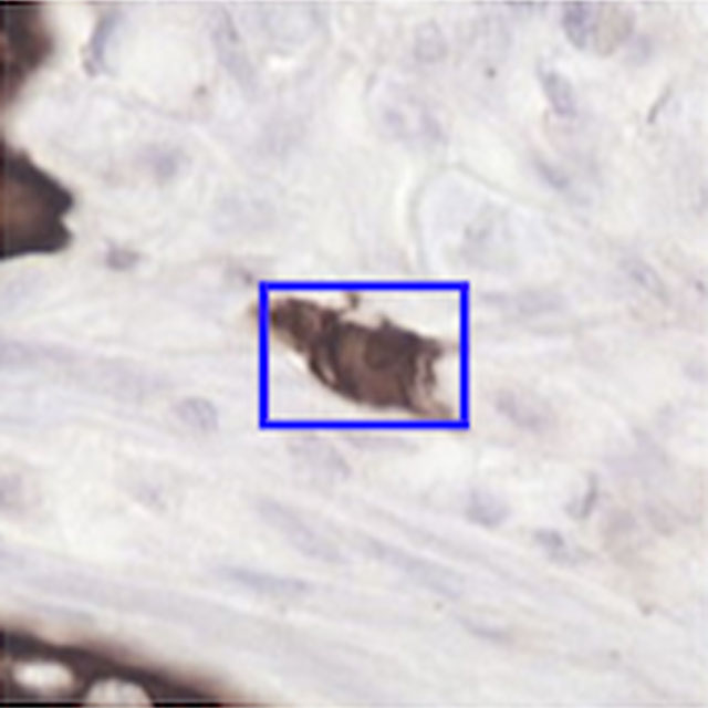
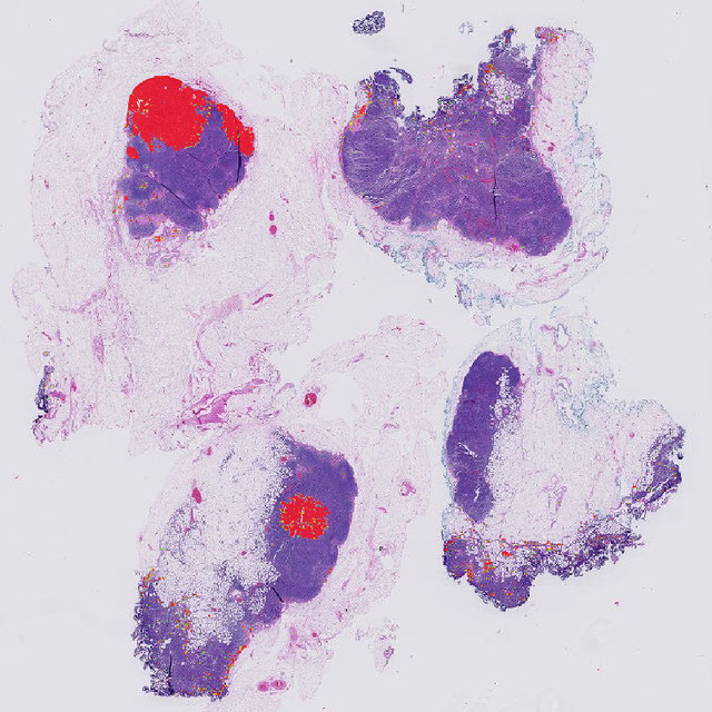
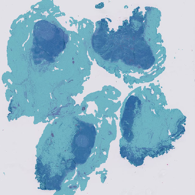
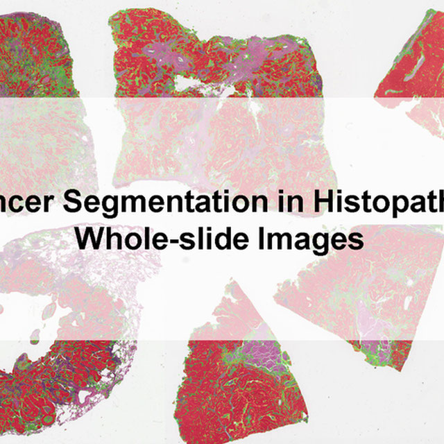
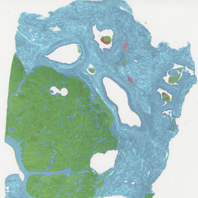
PAIP2020
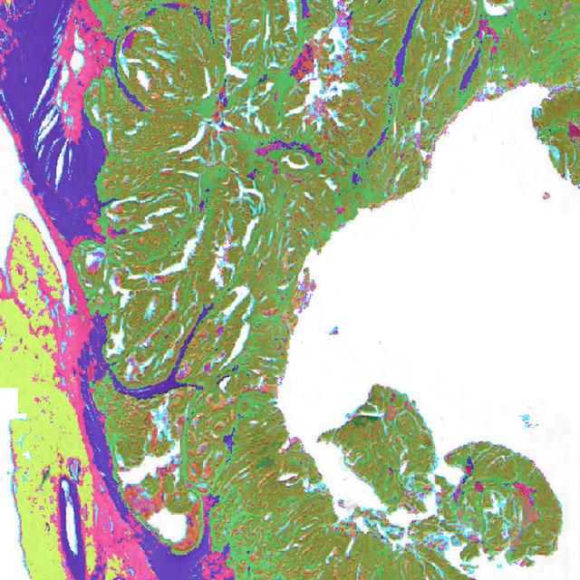
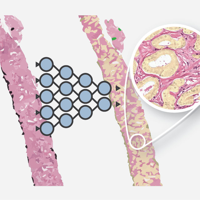
Challenge UserBuilt on the success of its predecessor, PAIP2020 is the second challenge organized by the Pathology AI Platform (PAIP) and the Seoul National University Hospital (SNUH). PAIP2020 will proceed to not only detect whole tumor areas in colorectal cancers but also to classify their molecular subtypes, which will lead to characterization of their heterogeneity with respect to prognoses and therapeutic responses. All participants should predict one of the molecular carcinogenesis pathways, i.e., microsatellite instability(MSI) in colorectal cancer, by performing digital image analysis without clinical tests. This task has a high clinical relevance as the currently used procedure requires an extensive microscopic assessment by pathologists. Therefore, those automated algorithms would reduce the workload of pathologists as a diagnostic assistance.


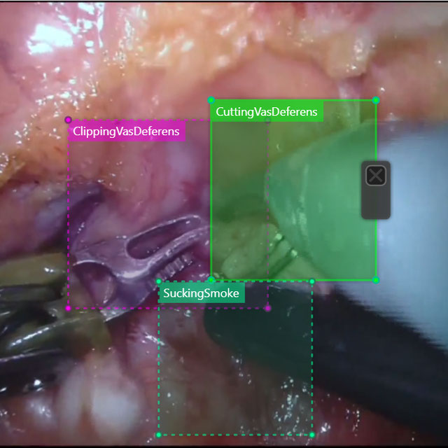
SARAS-MESAD
Challenge UserThis challenge is organized under MICCAI 2021, the 24th International Conference on Medical Image Computing and Computer Assisted Intervention. The event will be held from September 27th to October 1st 2021 in Strasbourg, France. The challenge focuses on multi-domain surgeon action detection.
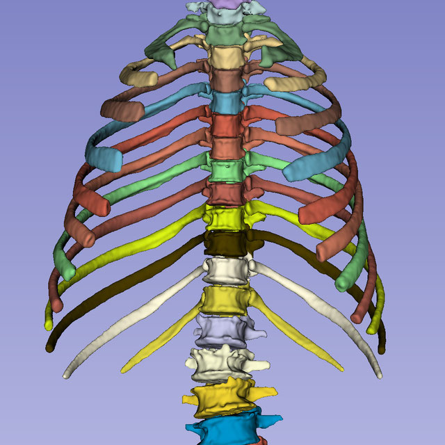
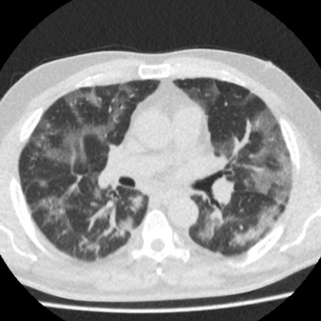
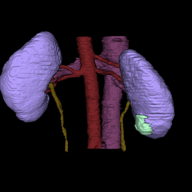
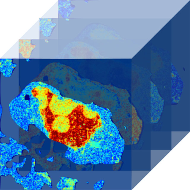
Parse2022
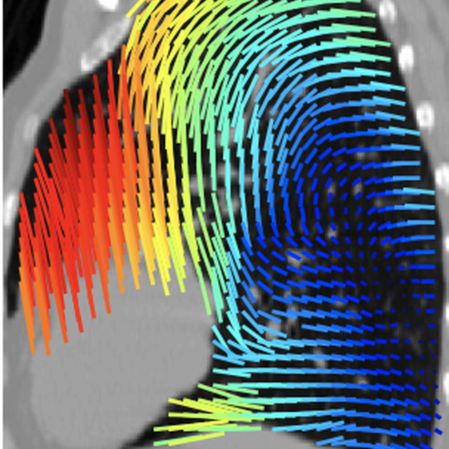
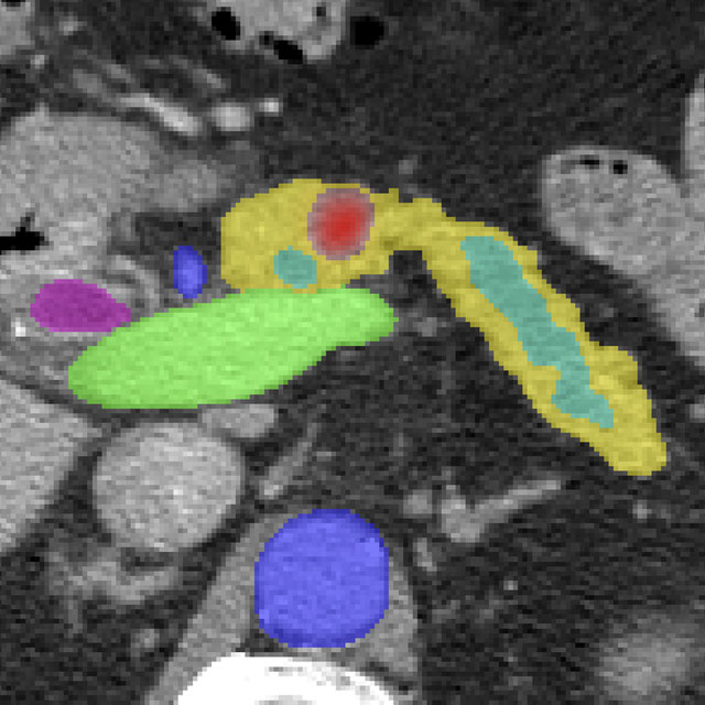
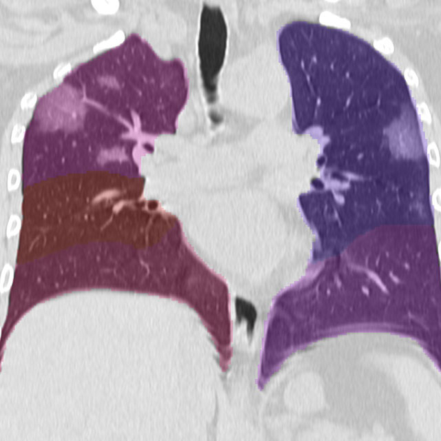
Challenge UserIt is of significant clinical interest to study pulmonary artery structures in the field of medical image analysis. One prerequisite step is to segment pulmonary artery structures from CT with high accuracy and low time-consuming. The segmentation of pulmonary artery structures benefits the quantification of its morphological changes for diagnosis of pulmonary hypertension and thoracic surgery. However, due to the complexity of pulmonary artery topology, automated segmentation of pulmonary artery topology is a challenging task. Besides, the open accessible large-scale CT data with well labeled pulmonary artery are scarce (The large variations of the topological structures from different patients make the annotation an extremely challenging process). The lack of well labeled pulmonary artery hinders the development of automatic pulmonary artery segmentation algorithm. Hence, we try to host the first Pulmonary ARtery SEgmentation challenge in MICCAI 2022 (Named Parse2022) to start a new research topic.
3D Teeth Scan Segmentation and Labeling Challenge MICCAI2022
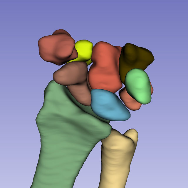
Challenge UserComputer-aided design (CAD) tools have become increasingly popular in modern dentistry for highly accurate treatment planning. In particular, in orthodontic CAD systems, advanced intraoral scanners (IOSs) are now widely used as they provide precise digital surface models of the dentition. Such models can dramatically help dentists simulate teeth extraction, move, deletion, and rearrangement and therefore ease the prediction of treatment outcomes. Although IOSs are becoming widespread in clinical dental practice, there are only few contributions on teeth segmentation/labeling available in the literature and no publicly available database. A fundamental issue that appears with IOS data is the ability to reliably segment and identify teeth in scanned observations. Teeth segmentation and labelling is difficult as a result of the inherent similarities between teeth shapes as well as their ambiguous positions on jaws.

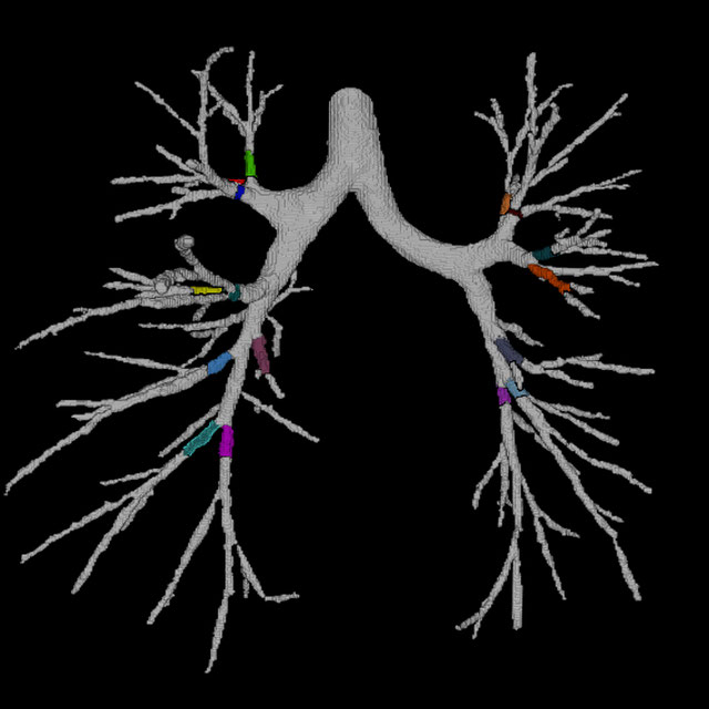
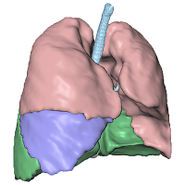
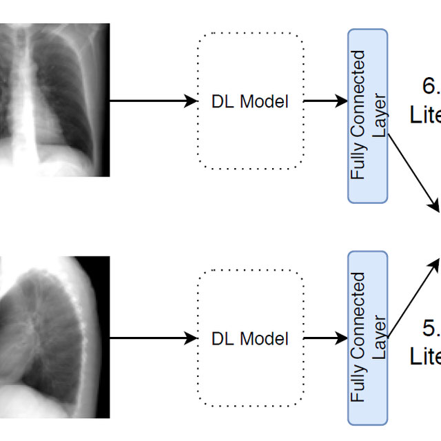
Multi-site, Multi-Domain Airway Tree Modeling (ATM’22)
Challenge UserAirway segmentation is a crucial step for the analysis of pulmonary diseases including asthma, bronchiectasis, and emphysema. The accurate segmentation based on X-Ray computed tomography (CT) enables the quantitative measurements of airway dimensions and wall thickness, which can reveal the abnormality of patients with chronic obstructive pulmonary disease (COPD). Besides, the extraction of patient-specific airway models from CT images is required for navigatiisted surgery.
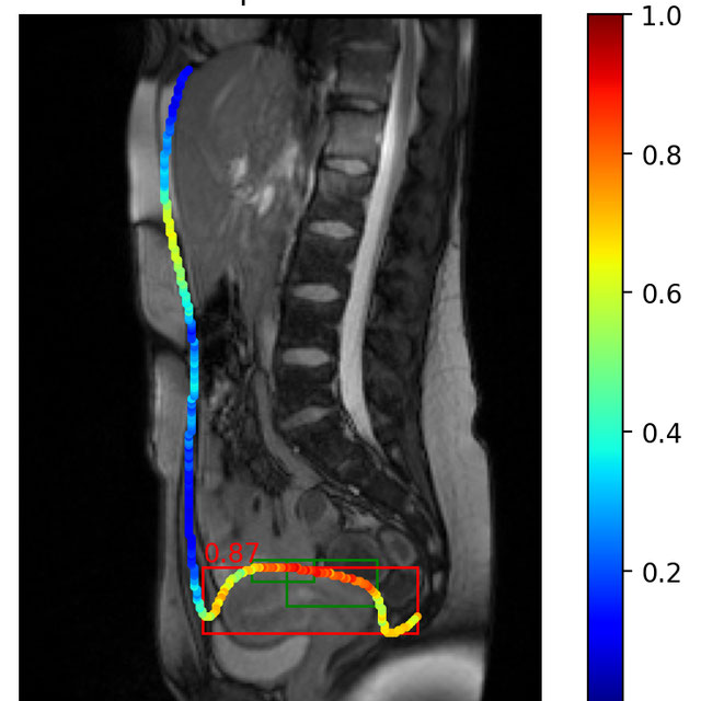
ToothFairy: Cone-Beam Computed Tomography Segmentation Challenge
Challenge EditorThis is the first edition of the ToothFairy challenge organized by the University of Modena and Reggio Emilia with the collaboration of Raudboud University. This challenge aims at pushing the development of deep learning frameworks to segment the Inferior Alveolar Canal (IAC) by incrementally extending the amount of publicly available 3D-annotated Cone Beam Computed Tomography (CBCT) scans. CBCT modality is becoming increasingly important for treatment planning and diagnosis in implant dentistry and maxillofacial surgery. The three-dimensional information acquired with CBCT can be crucial to plan a vast number of surgical interventions with the aim of preserving noble anatomical structures such as the Inferior Alveolar Canal (IAC), which contains the homonymous nerve (Inferior Alveolar Nerve, IAN). Deep learning models can support medical personnel in surgical planning procedures by providing a voxel-level segmentation of the IAN automatically extracted from CBCT scans.

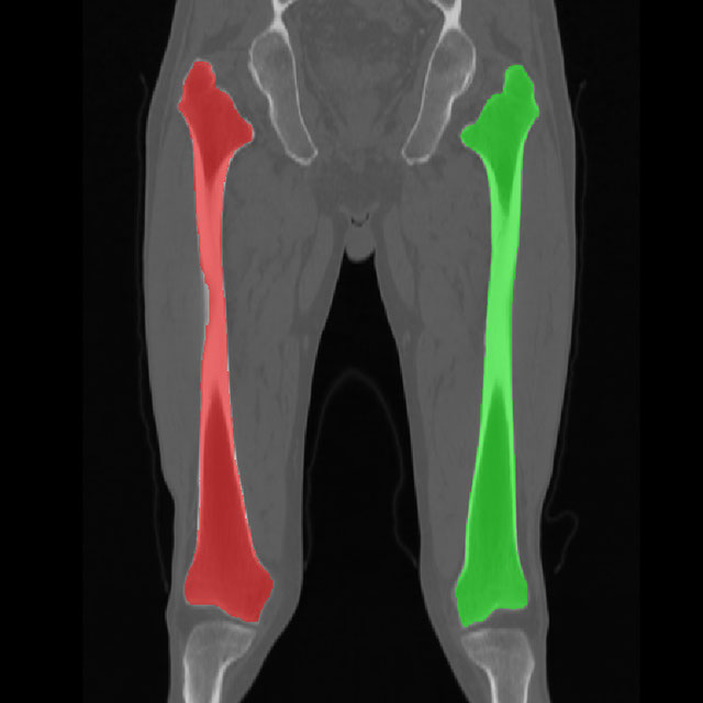
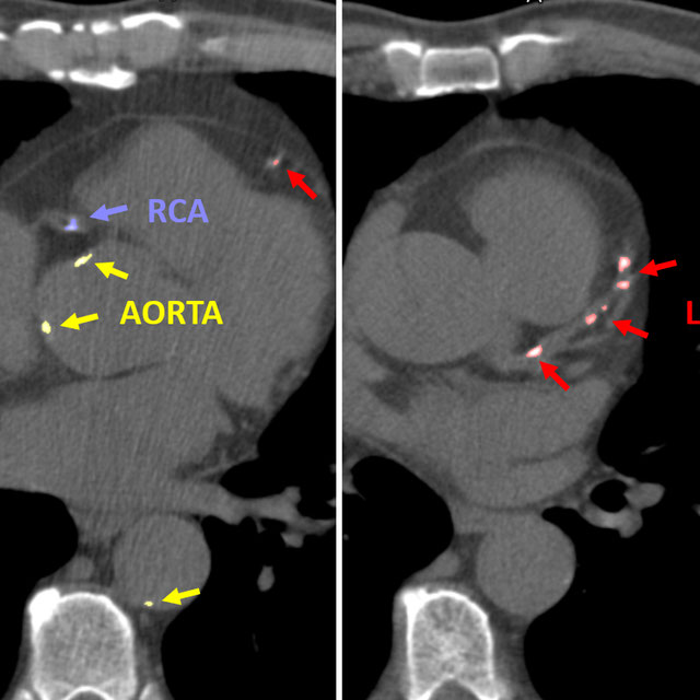
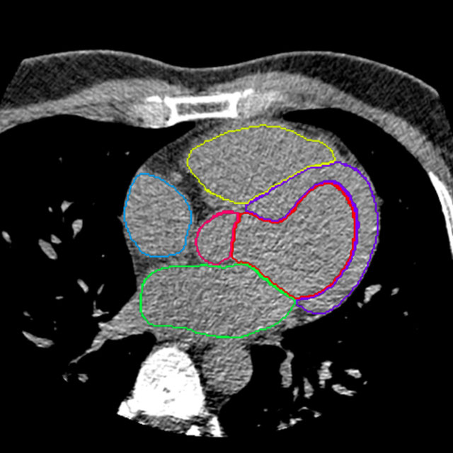
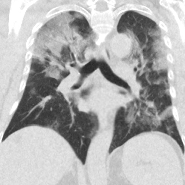
Whole-heart segmentation in non-contrast-enhanced CT
Algorithm UserAlgorithm for the automatic segmentation of cardiac structures in non-contrast-enhanced CT images. The structures to be segmented are the left ventricular myocardium, left ventricular cavity, right ventricle, left atrium, right atrium, ascending aorta, and the pulmonary artery trunk until the first bifurcation. The underlying publication can be found under https://doi.org/10.1002/mp.14451