Diagnostic Image Analysis Group
The Diagnostic Image Analysis Group is part of the Departments of Imaging, Pathology, Ophthalmology, and Radiation Oncology of Radboud University Medical Center. We develop computer algorithms to interpret and process medical images.
- Location
- Netherlands
- Website
- https://www.diagnijmegen.nl
- Editors
 koopman.t
 harm.van.zeeland
 pkcakeout
 siemdejong
 dnschouten
 MiriamGroeneveld
 chrisvanrun
 amickan
 KhrystynaFaryna
Activity Overview
RadboudCOVID
Archive
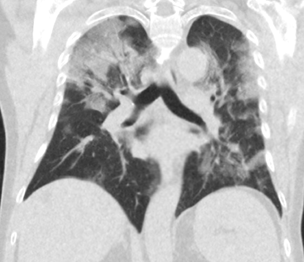
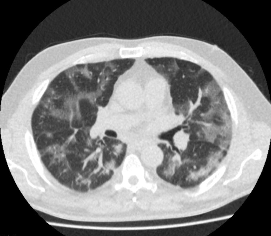
Data from RadboudUMC from Covid-19 (suspected) subjects
LOLA11
Archive
55 scans from the LOLA11 challenge
LUNA16
Archive
888 CT scans from the LUNA16 challenge
CORADS Score Exam
Reader Study
Assign a CORADS score to 25 cases. You will receive the results of the test by e-mail.
CORADS Score Practice
Reader Study
Practice CORADS scoring with 50 cases. You get instant feedback after every case.
Project AIR - DEMO - Lung nodule detection X-ray
Reader Study
Demonstration reader study to explore what it means to be a reader for Project AIR
Adhesion cine-MRI tutorial
Reader Study
A short 4-case tutorial for adhesion detection on abdominal cine-MRI
Demo educational reader study
Reader Study
Reader study for demo purposes
The PANORAMA Challenge - Reading Interface and Workflow Demo
Reader Study
PANORAMA is an all-new grand challenge with +2000 of carefully curated abdominal CECT exams, and tools to upload, run and validate AI in a secure environment, using standardized consensus-based guidelines.
LOLA11
Challenge
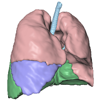
The goal of LOLA11 (LObe and Lung Analysis 2011) is to compare methods for (semi-)automatic segmentation of the lungs and lobes from chest computed tomography scans. Any team, whether from academia or industry, can join.
LUNA16
Challenge
The LUNA16 challenge: automatic nodule detection on chest CT
EMPIRE10
Challenge
Challenge for intra-subject registration of chest CT images.
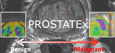
PROSTATEx
Challenge
Classification of clinical significance of prostate lesions using multi-parametric MRI data
HC18
Challenge
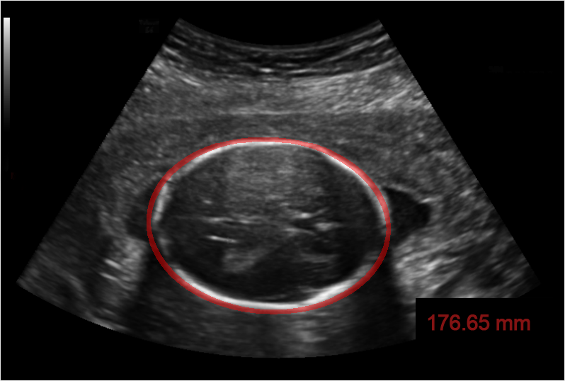
Automated measurement of fetal head circumference using 2D ultrasound images
NODE21
Challenge
NODE21: generate and detect nodules on chest radiographs
TIGER
Challenge
Grand challenge on automate assessment of tumor infiltrating lymphocytes in digital pathology slides of triple negative and Her2-positive breast cancers
The PI-CAI Challenge
Challenge
Artificial Intelligence and Radiologists at Prostate Cancer Detection in MRI
AIROGS
Challenge
Artificial Intelligence for RObust Glaucoma Screening Challenge
Project AIR - commercial AI for bone age prediction on hand XR
Challenge
Head-to-head performance evaluation of commercially available AI products. This challenge shows the results for bone age prediction on hand radiographs on a multicenter dataset (seven centers) from the Netherlands.
Project AIR - commercial AI for lung nodule detection on CXR
Challenge
Head-to-head performance evaluation of commercially available AI products. This challenge shows the results for lung nodule detection on chest radiographs on a multicenter dataset (seven centers) from the Netherlands.
Universal Lesion Segmentation Challenge '23
Challenge
Diagnostic Report Analysis: General Optimization of NLP
Challenge
Abdominal Circumference Operator-agnostic UltraSound measurement
Challenge
The LEOPARD Challenge
Challenge
MONKEY challenge: Detection of inflammation in kidney biopsies
Challenge
MONKEY (Machine-learning for Optimal detection of iNflammatory cells in KidnEY)
UNICORN
Challenge
Grand challenge on benchmarking vision-language foundation models in the digital pathology and radiology domain
PANTHER Challenge
Challenge
REport Generation in pathology using Pan-Asia Giga-pixel WSIs
Challenge
This project focuses on advancing automated pathology report generation using vision-language foundation models. It addresses the limitations of traditional NLP metrics (e.g., BLEU, METEOR, ROUGE) by emphasizing clinically relevant evaluation. The initiative includes standardized datasets, expert comparisons, and medical-domain-specific metrics to assess model performance. It also explores the integration of generated reports into diagnostic workflows with clinical feedback. To support fairness and generalizability, the challenge dataset comprises ~20,500 cases from six medical centers in Korea, Japan, India, Turkey, and Germany, promoting multicultural and multiethnic medical AI development.
BEETLE
Challenge
BEETLE is a multicenter, multiscanner benchmark for breast cancer histopathology segmentation. It focuses on multiclass semantic segmentation of hematoxylin and eosin (H&E)-stained whole-slide images (WSIs) into four tissue categories: invasive epithelium, non-invasive epithelium, necrosis, and other. The evaluation set comprises 170 densely annotated regions from 54 WSIs, covering all molecular subtypes and histological grades, thereby capturing much of the morphological heterogeneity seen in clinical practice. BEETLE provides a standardized resource for benchmarking breast cancer segmentation models, supporting the development of robust, generalizable algorithms for large-scale biomarker quantification across heterogeneous patient cohorts.
Lung cancer risk estimation on thorax CT scans - DSB2017 grt123
Algorithm
Automatic lung cancer risk estimation from thoracic CT scans
Spleen Segmentation
Algorithm
Automatic spleen segmentation on thorax-abdomen CT scans.
Vertebra segmentation and labeling
Algorithm
Segmentation and labeling of the vertebrae in CT scans with arbitrary field of view.
Tumor Detection in Lymph Nodes
Algorithm
CXR Cardiomegaly Detection
Algorithm
Detect cardiomegaly on chest radiographs through the segmentation of the heart and lungs .
Gleason Grading of Prostate Biopsies
Algorithm
Automated Gleason grading of prostate biopsies following the Gleason Grade Group system.
Segmentation of advanced AMD biomarkers in OCT
Algorithm
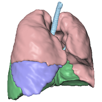
Pulmonary Lobe Segmentation
Algorithm
Automatic segmentation of pulmonary lobes on CT scans for patients with COPD or COVID-19.
Scaphoid fracture detection
Algorithm
Automatic detection of scaphoid fractures on hand, wrist, and scaphoid x-rays.
CORADS-AI
Algorithm
Segments pulmonary lobes and lesions and computes the CORADS and CT Severity Score from a non-contrast CT scan.
Pulmonary Nodule Malignancy Prediction
Algorithm
Deep Learning for Malignancy Risk Estimation of Low-Dose Screening CT Detected Pulmonary Nodules
HookNet-Breast
Algorithm
Segmentation algorithm for histopathology breast tissue.
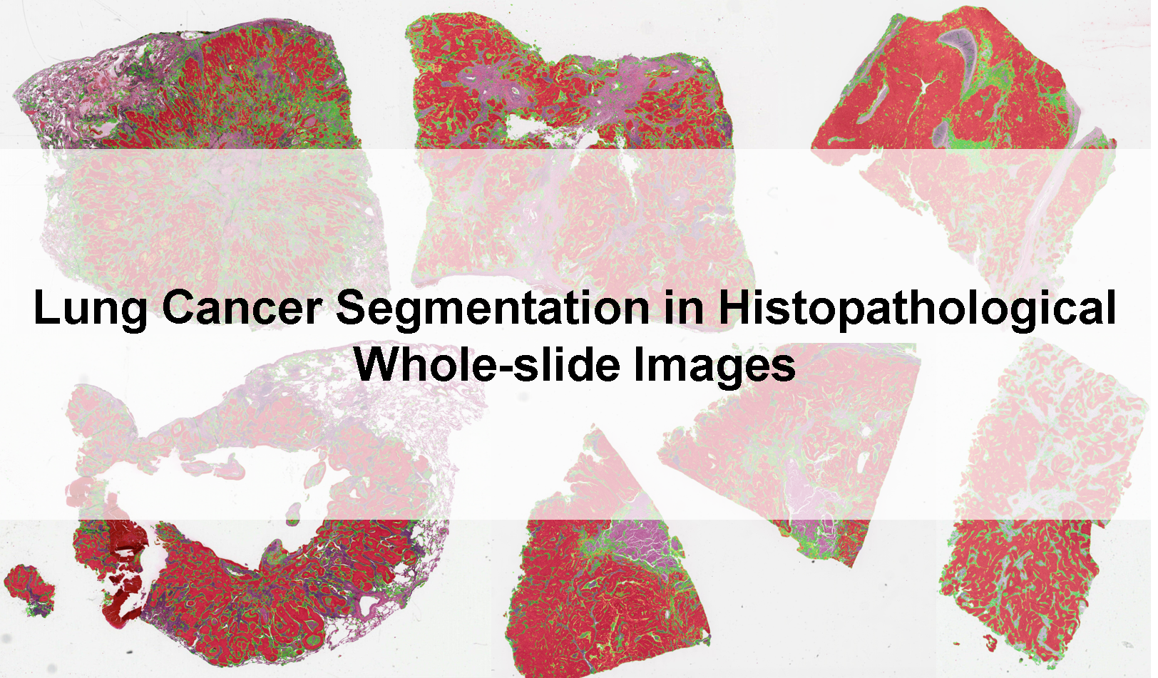
Lung Cancer Segmentation
Algorithm
Lung cancer segmentation in H&E stained histopathological images.
Lung cancer risk estimation on thorax CT scans - DSB2017 JulianDaniel
Algorithm
Automatic lung cancer risk estimation from thoracic CT scans
Fluid Segmentation in Retinal Optical Coherence Tomography (OCT)
Algorithm
Segments intraretinal fluid, subretinal fluid, and pigment epithelial detachments in Optical Coherence Tomography scans. Optimized for Spectralis, Cirrus and Topcon scanners.
HookNet-Lung
Algorithm
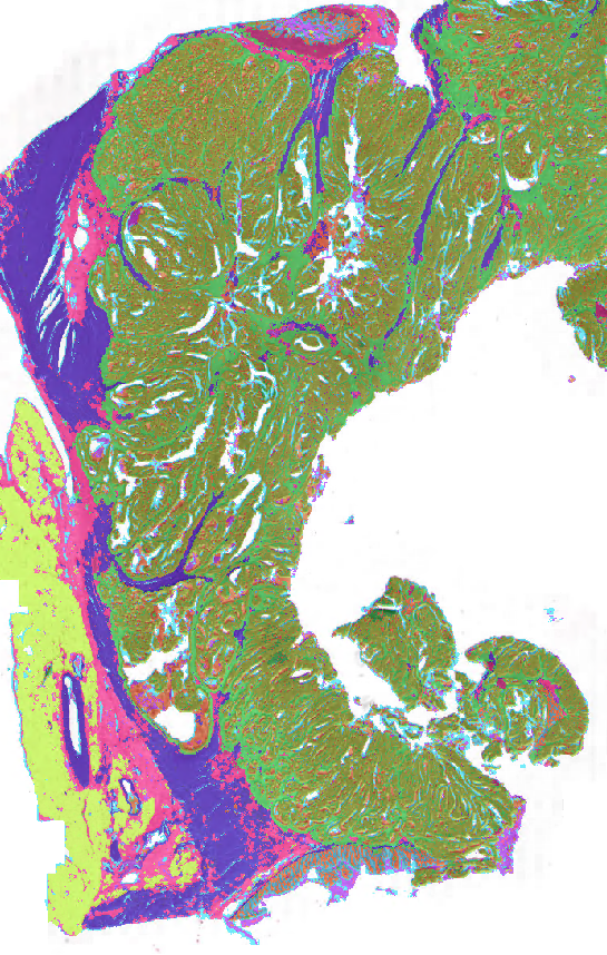
Segmentation algorithm for histopathology lung tissue.
Femur segmentation in CT
Algorithm
Segments the left and right femur in CT images
Neural Image Compression
Algorithm
Compresses whole slide images into much smaller volumes
CXR Total Lung Volume Measurement
Algorithm
Measurement of total lung volume from chest radiographs using frontal and lateral radiographs
Clinically Significant Prostate Cancer Detection in bpMRI
Algorithm
Deep learning-based 3D detection/diagnosis model trained, validated and tested using 2732 prostate biparametric MRI exams from two centers.
Demo Vessel Segmentation
Algorithm
Demonstrates a vessel segmentation algorithm.
Colon Tissue segmentation
Algorithm
Tissue segmentation network for colon histopathology images
Body composition
Algorithm
Locates L3 in a CT scan and measures the area of subcutaneous, visceral, and intermuscular adipose tissue and smooth muscle. The average Hounsfield Units of each area are also computed.
Nuclear Pleomorphism Scoring
Algorithm
Scoring nuclear pleomorphism grade in whole-slide breast histopathology images
Hip segmentation in CT
Algorithm
Segments the left and right hip bones in CT images
Vertebral Fracture Assessment
Algorithm
A neural network that assesses vertebral fractures according to the Genant classification
Vertebral Abnormality Scoring
Algorithm
Score from 0 to 100 that expresses how abnormal the shape of a vertebra is
Lobe-Wise Lung Function Estimation from CT
Algorithm
Produces patient-level and lobe-level estimates of DLCO and of FEV1 and FVC pre- and post-bronchodilator
STOIC2021 baseline
Algorithm
Example algorithm for the STOIC2021 COVID-19 AI Challenge
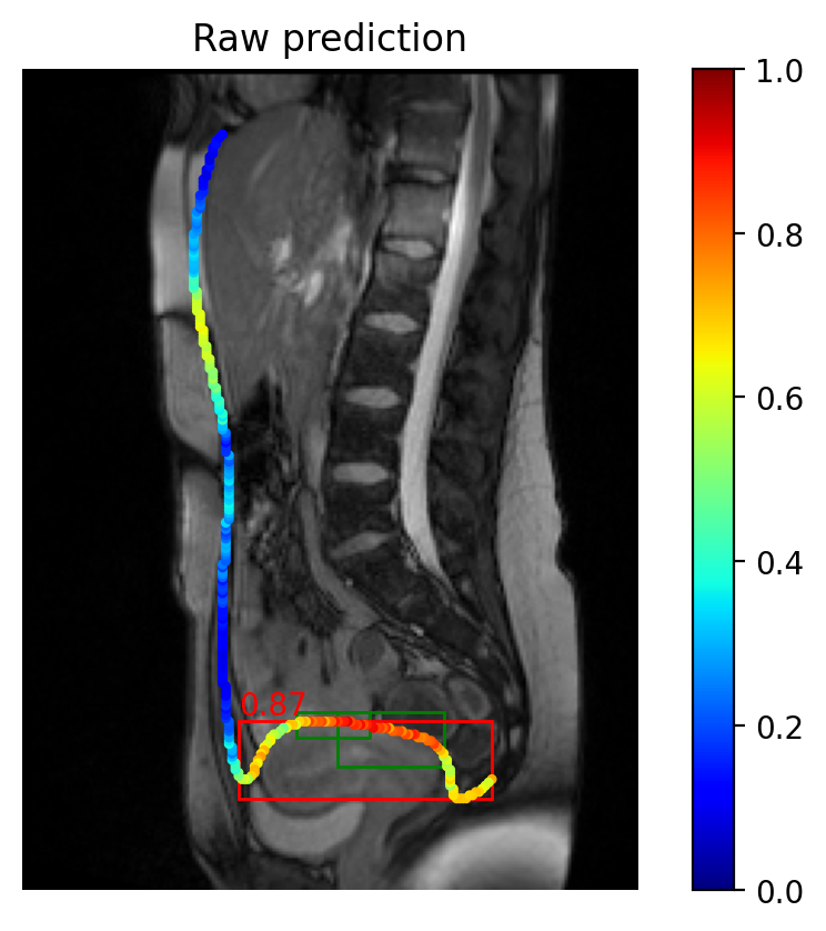
Visceral slide on abdominal cine-MRI
Algorithm
This algorithm calculates the visceral slide along the contour of the abdominal cavity, using segmentation and registration.
Breast Cancer Segmentation and Scoring in H&E
Algorithm
Liver segmentation
Algorithm
Segments the liver and liver tumors using nnUNet
Multi-view scaphoid fracture detection
Algorithm
Automated scaphoid fracture detection on conventional radiographs of the hand, wrist, and scaphoid in any view.
Lung nodule detection for routine clinical CT scans
Algorithm
Deep learning for the detection of pulmonary nodules in chest CT scans
AIROGS Baseline
Algorithm
A baseline algorithm for the AIROGS challenge
Clinically Significant Prostate Cancer Detection in bpMRI using models trained with Report Guided Annotations
Algorithm
Airway Anatomical Labeling
Algorithm
Given an airway segmentation where individual airway branches are extracted, this algorithm will automatically find 18 segmental branches, including 8 from the left lung (LB1+2, LB3, LB4, LB5, LB6, LB7+8, LB9, and LB10) and 10 from the right lung (RB1-10).
Prostate Segmentation
Algorithm
Whole-Gland Prostate Segmentation in bpMRI
Tiger Algorithm Example
Algorithm
example on how to create an algorithm for the tiger challenge
Age-related macular degeneration (AMD) Staging in Optical Coherence Tomography (OCT) with UBIX for Increased Reliability
Algorithm
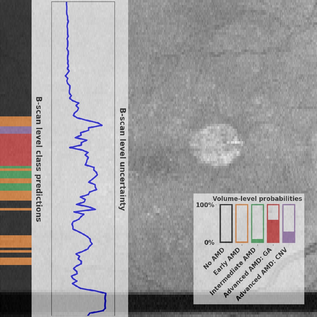
Deep learning models for optical coherence tomography (OCT) classification often perform well on data from scanners that were also used during training. However, when these models are applied to data from different vendors, their performance tends to drop substantially. Artifacts that only occur within scans from specific scanners are major causes of this poor generalizability. We aimed to improve this generalizability of deep learning models for OCT classifi- cation. To reduce the effect of vendor-specific artifacts, we propose Uncertainty-Based Instance eXclusion (UBIX), of which we define a hard and a soft variant. UBIX aims to suppress the contributions of B-scans with unseen artifacts to the final OCT-level outputs. Suppression is based on out-of-distribution detection of B-scans, which are instances in our multiple instance learning approach.
Breast and fibroglandular tissue segmentation
Algorithm
PI-CAI: Baseline nnU-Net (supervised)
Algorithm
Baseline algorithm submission for PI-CAI based on the nnU-Net framework
Wrist segmentation
Algorithm
Segments the carpal bones and radius and ulna in (dynamic) CT scans of the wrist.
Tibia segmentation in CT
Algorithm
Segments the left and right tibia in CT images
Rib segmentation
Algorithm
Segments and labels the ribs in CT images
PI-CAI: Baseline nnDetection (supervised)
Algorithm
Baseline algorithm submission for PI-CAI based on the nnDetection framework
PI-CAI: Baseline nnU-Net (semi-supervised)
Algorithm
Baseline semi-supervised algorithm submission for PI-CAI based on the nnU-Net framework
PI-CAI: Baseline U-Net (semi-supervised)
Algorithm
PI-CAI: Baseline nnDetection (semi-supervised)
Algorithm
Baseline semi-supervised algorithm submission for PI-CAI based on the nnDetection framework
Endometrial Carcinoma classification
Algorithm
CLAM model that computes the probability of the WSI being (pre)malignant and also outputs an interpretable heatmap.
Colon Budding in IHC
Algorithm
Automatic tumor bud detection in IHC stained slides of CRC
Subsolid nodule segmentation
Algorithm
Algorithm for segmenting sub-solid pulmonary nodules in CT
PI-CAI: Baseline nnU-Net (supervised) trained on PI-CAI: Private and Public Training Dataset
Algorithm
Lymphocytes detection in immunohistochemistry
Algorithm
JawFracNet
Algorithm
Segment mandible and mandibular fractures in head CBCT scan
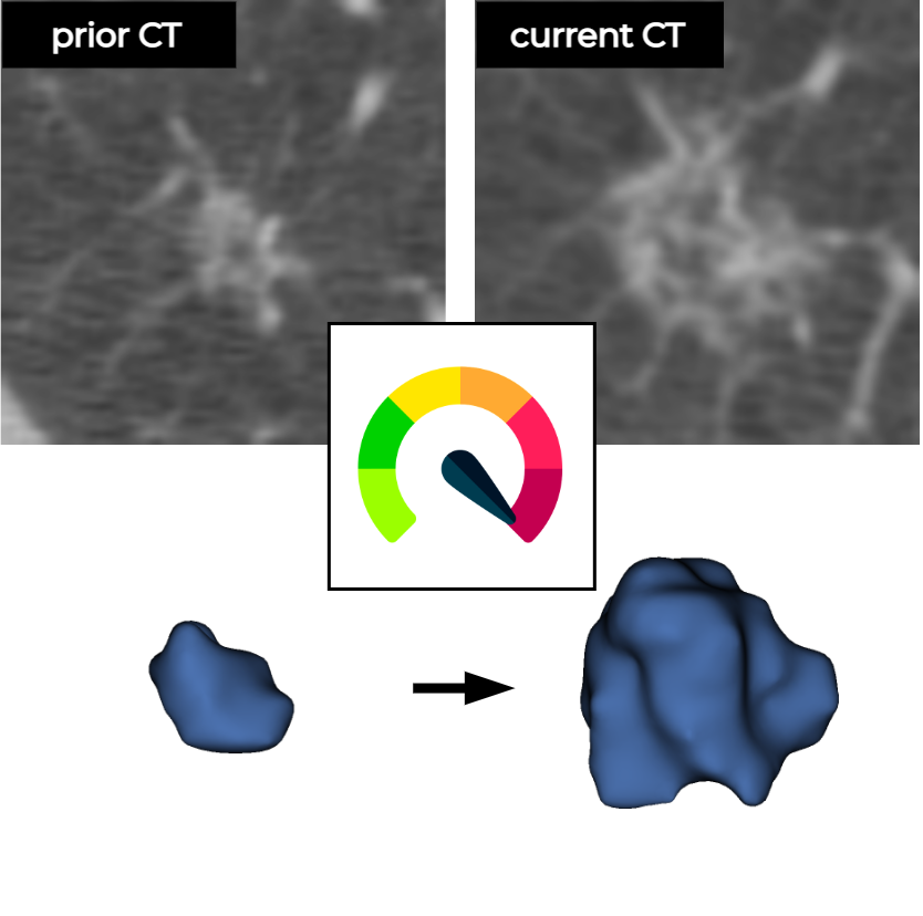
Deep learning to estimate pulmonary nodule malignancy risk using a current and a prior CT image
Algorithm
Deep learning to estimate pulmonary nodule malignancy risk using a prior CT image
PI-CAI: Baseline nnU-Net (semi-supervised) trained on PI-CAI: Private and Public Training Dataset
Algorithm
HookNet-TLS
Algorithm
A model for detection of TLS and GC in histopathology.
PI-CAI: Baseline nnDetection (semi-supervised) trained on PI-CAI: Private and Public Training Dataset
Algorithm
PI-CAI: Baseline U-Net (supervised) trained on PI-CAI: Private and Public Training Dataset
Algorithm
PI-CAI: Baseline U-Net (semi-supervised) trained on PI-CAI: Private and Public Training Dataset
Algorithm
PI-CAI: Baseline nnDetection (supervised) trained on PI-CAI Private and Public Training Dataset
Algorithm
PI-CAI: Baseline nnDetection (supervised) trained on PI-CAI Private and Public Training Dataset
SPIDER Baseline nnU-Net
Algorithm
Carpal instability measurements
Algorithm
Automated AI pipeline for conducting carpal instability measurements on conventional radiographs of the hand and wrist.
SPIDER Baseline IIS
Algorithm
Tumor proportion score in non-small cell lung cancer
Algorithm
Automatic quantification of the tumor proportion score in NSCLC histology
Nuclei detection in immunohistochemistry
Algorithm
Automatic detection of nuclei (hematoxylin positive) in immunohistochemistry WSIs
Neural ODEs for Segmentation of Colon Glands
Algorithm
Neural Ordinary Differential Equations for Semantic Segmentation of Individual Colon Glands
DRAGON DistilBERT Base General-domain
Algorithm
DRAGON DistilBERT Base General-domain
DRAGON BERT Base General-domain
Algorithm
DRAGON BERT Base General-domain
DRAGON RoBERTa Base General-domain
Algorithm
DRAGON RoBERTa Base General-domain
DRAGON Longformer Base General-domain
Algorithm
DRAGON Longformer Base General-domain
DRAGON Longformer Large General-domain
Algorithm
DRAGON Longformer Large General-domain
DRAGON RoBERTa Large General-domain
Algorithm
DRAGON RoBERTa Large General-domain
PDAC Tumor Segmentation
Algorithm
This algorithm takes an HE histology images of the pancreas, segments the tissue, segments the epithelium and segments the tumor (if present)
Universal Lesion Segmentation [ULS23 Baseline]
Algorithm
Univeral Lesion Segmentation algorithm for Computed Tomography
Airway nodule detection for routine clinical CT scans
Algorithm
Deep learning for the detection of airway nodules in chest CT scans
DRAGON BERT Base Mixed-domain
Algorithm
DRAGON BERT Base Mixed-domain
DRAGON RoBERTa Base Mixed-domain
Algorithm
DRAGON RoBERTa Base Mixed-domain
DRAGON RoBERTa Large Mixed-domain
Algorithm
DRAGON RoBERTa Large Mixed-domain
DRAGON Longformer Base Mixed-domain
Algorithm
DRAGON Longformer Base Mixed-domain
DRAGON Longformer Large Mixed-domain
Algorithm
DRAGON Longformer Large Mixed-domain
DRAGON BERT Base Domain-specific
Algorithm
DRAGON BERT Base Domain-specific
DRAGON RoBERTa Base Domain-specific
Algorithm
DRAGON RoBERTa Base Domain-specific
DRAGON Longformer Base Domain-specific
Algorithm
DRAGON Longformer Base Domain-specific
DRAGON Longformer Large Domain-specific
Algorithm
DRAGON Longformer Large Domain-specific
DRAGON CLTL MedRoBERTa.nl
Algorithm
DRAGON CLTL MedRoBERTa.nl Domain-specific
DRAGON RoBERTa Large Domain-specific V2
Algorithm
DRAGON RoBERTa Large Domain-specific V2
Pancreatic tumor segmentation on T2W MRI :PANTHER challenge baseline
Algorithm
Baseline algorithm for Task 2 of the PANTHER challenge
PDAC tumor segmentation on T1W-CE-MRI :PANTHER challenge baseline
Algorithm
Baseline algorithm for Task 1 of the PANTHER challenge
PRISM Embedder
Algorithm
Extracts slide-level representations from whole-slide images using PRISM.
Kidney Tissue Segmentation
Algorithm
PTC Segmentation
Algorithm
This model segments the Peritubular Capillaries (PTCs) from the rest of the tissue on PAS-stained WSI's of kindey transplant biopsies.
Deep Learning Image Quality for Prostate MRI
Algorithm


